9VX3 | pdb_00009vx3

Crystal structure of the peptide-bound form of HisMab-1 Fv-clasp


Experimental Data Snapshot

  • Method: X-RAY DIFFRACTION
  • Resolution: 2.39 Å
  • R-Value Free: 
    0.237 (Depositor), 0.239 (DCC) 
  • R-Value Work: 
    0.196 (Depositor), 0.196 (DCC) 
  • R-Value Observed: 
    0.198 (Depositor) 

Starting Models: experimental
View more details

wwPDB Validation   3D Report Full Report


This is version 1.1 of the entry. See complete history


Literature

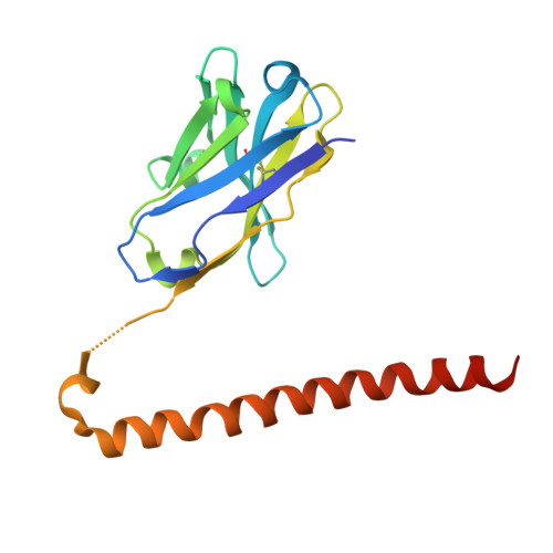
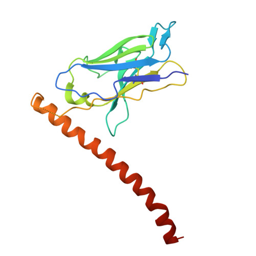
Functional and Structural Characterization of a Novel Anti-His-tag Antibody, HisMab-1.

Hitomi, N.Hoshi, S.Kaneko, M.K.Kato, R.Iwasaki, K.Takagi, J.Kato, Y.Harada-Hikita, A.Arimori, T.

(2025) J Mol Biology 438: 169574-169574

  • DOI: https://doi.org/10.1016/j.jmb.2025.169574
  • Primary Citation of Related Structures:  
    9VX2, 9VX3

  • PubMed Abstract: 

    The polyhistidine tag (His-tag) is one of the most widely used peptide tags for the purification of recombinant proteins, owing to its compatibility with immobilized metal affinity chromatography. While numerous anti-His-tag antibodies are commercially available, their quantitative affinity data and structural insights are limited. Here, we present a detailed physicochemical and structural characterization of a novel anti-His-tag antibody, HisMab-1. Isothermal titration calorimetry showed that the Fab fragment of HisMab-1 binds to a hexahistidine peptide in an enthalpy-driven manner, with a dissociation constant (K D ) of ∼30 nM at a neutral pH. The crystal structure of the HisMab-1-hexahistidine peptide complex at 2.39-Å resolution revealed that HisMab-1 primarily recognizes the first, second, fourth, and fifth histidine residues of the peptide through multiple interactions, including hydrogen bonding and π-π stacking, which collectively contribute to the high specificity of the antibody. Notably, HisMab-1 also binds to a His-tag embedded within a conformationally constrained β-hairpin loop without reducing affinity, highlighting its structural adaptability. These findings establish HisMab-1 as a high-affinity, high-specificity, structurally validated anti-His-tag antibody with broad potential in diverse protein engineering and structural biology applications.


  • Organizational Affiliation
    • Laboratory for Protein Synthesis and Expression, Institute for Protein Research, The University of Osaka, 3-2, Yamadaoka, Suita, Osaka 565-0871, Japan.

Macromolecules
Find similar proteins by:  (by identity cutoff)  |  3D Structure
Entity ID: 1
MoleculeChains Sequence LengthOrganismDetailsImage
HisMab-1VH(S112C),Serine/threonine-protein kinase 4 18kDa subunit
A, D
175Mus musculusHomo sapiens
This entity is chimeric
Mutation(s): 0 
EC: 2.7.11.1
UniProt & NIH Common Fund Data Resources
Find proteins for Q13043 (Homo sapiens)
Explore Q13043 
Go to UniProtKB:  Q13043
PHAROS:  Q13043
GTEx:  ENSG00000101109 
Entity Groups  
Sequence Clusters30% Identity50% Identity70% Identity90% Identity95% Identity100% Identity
UniProt GroupQ13043
Sequence Annotations
Expand
  • Reference Sequence
Find similar proteins by:  (by identity cutoff)  |  3D Structure
Entity ID: 2
MoleculeChains Sequence LengthOrganismDetailsImage
HisMab-1VL,Serine/threonine-protein kinase 4 18kDa subunit
B, E
167Mus musculusHomo sapiens
This entity is chimeric
Mutation(s): 1 
EC: 2.7.11.1
UniProt & NIH Common Fund Data Resources
Find proteins for Q13043 (Homo sapiens)
Explore Q13043 
Go to UniProtKB:  Q13043
PHAROS:  Q13043
GTEx:  ENSG00000101109 
Entity Groups  
Sequence Clusters30% Identity50% Identity70% Identity90% Identity95% Identity100% Identity
UniProt GroupQ13043
Sequence Annotations
Expand
  • Reference Sequence

Find similar proteins by:  Sequence   |   3D Structure  

Entity ID: 3
MoleculeChains Sequence LengthOrganismDetailsImage
Polyhistidine peptide
C, F
6synthetic constructMutation(s): 0 
Sequence Annotations
Expand
  • Reference Sequence
Experimental Data & Validation

Experimental Data

  • Method: X-RAY DIFFRACTION
  • Resolution: 2.39 Å
  • R-Value Free:  0.237 (Depositor), 0.239 (DCC) 
  • R-Value Work:  0.196 (Depositor), 0.196 (DCC) 
  • R-Value Observed: 0.198 (Depositor) 
Space Group: P 1 21 1
Unit Cell:
Length ( Å )Angle ( ˚ )
a = 54.139α = 90
b = 69.512β = 93.78
c = 93.396γ = 90
Software Package:
Software NamePurpose
PHENIXrefinement
XDSdata reduction
XDSdata scaling
PHASERphasing

Structure Validation

View Full Validation Report


Entry History & Funding Information

Deposition Data


Funding OrganizationLocationGrant Number
Japan Society for the Promotion of Science (JSPS)JapanJP21H02416
Japan Society for the Promotion of Science (JSPS)JapanJP25K02215
Japan Agency for Medical Research and Development (AMED)JapanJP25am0521010
Japan Agency for Medical Research and Development (AMED)JapanJP25ama121001
Japan Agency for Medical Research and Development (AMED)JapanJP25ama121011
Japan Agency for Medical Research and Development (AMED)JapanJP25ama121008

Revision History  (Full details and data files)

  • Version 1.0: 2025-12-17
    Type: Initial release
  • Version 1.1: 2025-12-24
    Changes: Database references