


Computational design of verteporfin binding protein
Tang, X., Li, Y.H., Xu, Y.L., Kou, L.Y., Ji, P.F.To be published.
Experimental Data Snapshot
Starting Model: in silico
View more details
wwPDB Validation 3D Report Full Report

Entity ID: 1 | |||||
|---|---|---|---|---|---|
| Molecule | Chains | Sequence Length | Organism | Details | Image |
| VTP-4 | 182 | synthetic construct | Mutation(s): 0 | 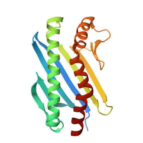 | |
| Ligands 2 Unique | |||||
|---|---|---|---|---|---|
| ID | Chains | Name / Formula / InChI Key | 2D Diagram | 3D Interactions | |
| P15 Download:Ideal Coordinates CCD File | D [auth A] | 2,5,8,11,14,17-HEXAOXANONADECAN-19-OL C13 H28 O7 FHHGCKHKTAJLOM-UHFFFAOYSA-N |  | ||
| EDO Download:Ideal Coordinates CCD File | B [auth A], C [auth A] | 1,2-ETHANEDIOL C2 H6 O2 LYCAIKOWRPUZTN-UHFFFAOYSA-N |  | ||
| Length ( Å ) | Angle ( ˚ ) |
|---|---|
| a = 35.606 | α = 90 |
| b = 58.488 | β = 90 |
| c = 83.828 | γ = 90 |
| Software Name | Purpose |
|---|---|
| REFMAC | refinement |
| XDS | data reduction |
| Aimless | data scaling |
| PHASER | phasing |
| Funding Organization | Location | Grant Number |
|---|---|---|
| National Natural Science Foundation of China (NSFC) | China | 22377107 |


