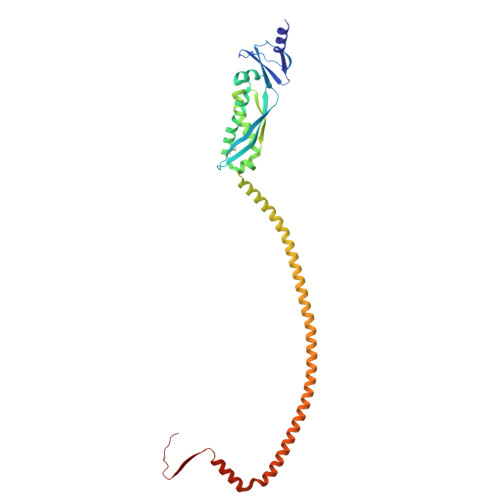
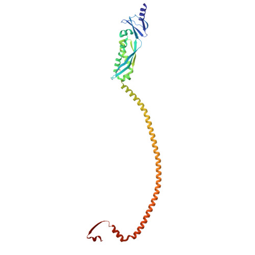
The SPFH (stomatin, prohibitin, flotillin, and HflK/C) family proteins are proposed scaffolds for organizing functional membrane microdomains (FMMs) on various cellular membranes. Erlin1 and Erlin2, two endoplasmic reticulum (ER)-residing SPFH members, as heteromeric complexes, participate in ER-associated protein degradation (ERAD). However, the mechanisms underlying Erlin-mediated FMM organization and ERAD regulation remain poorly understood. Here, through cryoelectron microscopy (cryo-EM), we find that the human Erlin1/2 complex forms a 26-mer cage assembly, defining a nanometer-sized microdomain on the luminal leaflet. The intramembrane region of each subunit constitutes a specific phosphatidylinositol-binding pocket. ER proteins can be recruited to both the interior and exterior of these cages. By caging cargoes, the Erlin1/2 complex physically secludes them from their substrates or binding partners, conferring another layer of regulation on their functions. Moreover, individual cages can cluster to organize FMMs of different sizes. These dynamic properties underscore a general regulatory role of Erlin1/2 in various ER-related biological processes, including coronaviral replication.
Organizational Affiliation:
State Key Laboratory of Membrane Biology, School of Life Sciences, Peking University, Beijing 100871, China.
Peking-Tsinghua Joint Center for Life Sciences, Academy for Advanced Interdisciplinary Studies, Peking University, Beijing 100871, China.
State Key Laboratory of Membrane Biology and Institute of Molecular Medicine, College of Future Technology, Peking University, Beijing 100871, China.
Peking-Tsinghua Joint Center for Life Sciences, Academy for Advanced Interdisciplinary Studies, Peking University, Beijing 100871, China; Center for Quantitative Biology, Academy for Advanced Interdisciplinary Studies, Peking University, Beijing 100871, China.
Peking-Tsinghua Joint Center for Life Sciences, Academy for Advanced Interdisciplinary Studies, Peking University, Beijing 100871, China; Changping Laboratory, Beijing 102206, China.
Changping Laboratory, Beijing 102206, China.
State Key Laboratory of Membrane Biology and Institute of Molecular Medicine, College of Future Technology, Peking University, Beijing 100871, China; National Biomedical Imaging Center, Peking University, Beijing 100871, China.
Peking-Tsinghua Joint Center for Life Sciences, Academy for Advanced Interdisciplinary Studies, Peking University, Beijing 100871, China; Center for Quantitative Biology, Academy for Advanced Interdisciplinary Studies, Peking University, Beijing 100871, China. Electronic address: c.song@pku.edu.cn.
Peking-Tsinghua Joint Center for Life Sciences, Academy for Advanced Interdisciplinary Studies, Peking University, Beijing 100871, China; State Key Laboratory of Membrane Biology and Institute of Molecular Medicine, College of Future Technology, Peking University, Beijing 100871, China; Beijing Advanced Center of RNA Biology (BEACON), Peking University, Beijing 100871, China. Electronic address: xiaowei_chen@pku.edu.cn.
State Key Laboratory of Membrane Biology, School of Life Sciences, Peking University, Beijing 100871, China; Peking-Tsinghua Joint Center for Life Sciences, Academy for Advanced Interdisciplinary Studies, Peking University, Beijing 100871, China; National Biomedical Imaging Center, Peking University, Beijing 100871, China; Changping Laboratory, Beijing 102206, China; Beijing Advanced Center of RNA Biology (BEACON), Peking University, Beijing 100871, China. Electronic address: gaon@pku.edu.cn.
AA [auth N], BA [auth O], CA [auth P], DA [auth Q], EA [auth R],
AA [auth N], BA [auth O], CA [auth P], DA [auth Q], EA [auth R], FA [auth S], GA [auth T], HA [auth U], IA [auth V], JA [auth W], KA [auth X], LA [auth Y], MA [auth Z], NA [auth n], OA [auth o], PA [auth p], QA [auth q], RA [auth r], SA [auth s], TA [auth t], UA [auth u], VA [auth v], WA [auth w], XA [auth x], YA [auth y], ZA [auth z]