9VQ2 | pdb_00009vq2

Cryo-EM structure of a human flippase mutant ATP11C Q79A-CDC50A in the PtdSer-occluded E2-AlF state


Experimental Data Snapshot

  • Method: ELECTRON MICROSCOPY
  • Resolution: 3.40 Å
  • Aggregation State: PARTICLE 
  • Reconstruction Method: SINGLE PARTICLE 

wwPDB Validation   3D Report Full Report


This is version 1.1 of the entry. See complete history


Literature

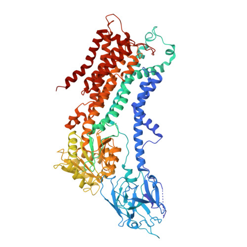
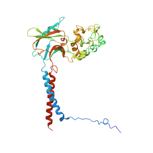
Cryo-EM structure of the ATP11C Q79E mutant reveals the structural basis for altered Phospholipid recognition.

Qian, Y.Gopalasingam, C.C.Gerle, C.Shigematsu, H.Abe, K.Oshima, A.

(2025) J Biological Chem 302: 110935-110935

  • DOI: https://doi.org/10.1016/j.jbc.2025.110935
  • Primary Citation of Related Structures:  
    9VKG, 9VNT, 9VQ2, 9VSL

  • PubMed Abstract: 

    Closely related P4-ATPases, ATP11A and ATP11C, act as major phospholipid flippases in the plasma membrane of mammalian cells, with strict substrate specificity for phosphatidylserine (PS) and phosphatidylethanolamine (PE), but not for phosphatidylcholine (PC), thereby contributing to the asymmetric distribution of PS and PE across bilayers. A previously reported disease-associated Q84E mutation in ATP11A confers the ability to flip PC, implicating the involvement of this conserved residue in substrate specificity. We performed cryo-EM analysis for the equivalent mutant Q79E of ATP11C to address the structural basis for its unusual substrate specificity. Measurement of ATPase activity revealed that the ATP11C Q79E mutant retained PS-dependent activity, whilst gaining robust PC-dependent activity, indicative of expanded substrate specificity, consistent with reported properties in ATP11A Q84E. The cryo-EM structure of ATP11C Q79E mutant in the PC-occluded E2-P i state revealed a PC molecule in a reshaped binding pocket. Due to the Q79E mutation and associated conformational changes in its surrounding residues, including Ser91and Asn352, the binding pocket has additional space to accommodate the bulky choline headgroup. Our results provide structural and functional insights into how a single point mutation can alter substrate specificity in a P4-ATPase.


  • Organizational Affiliation
    • Department of Chemistry, Faculty of Science, Hokkaido University, Japan; Graduate School of Pharmaceutical Sciences, Nagoya University, Nagoya, Japan.

Macromolecules
Find similar proteins by:  (by identity cutoff)  |  3D Structure
Entity ID: 1
MoleculeChains Sequence LengthOrganismDetailsImage
Phospholipid-transporting ATPase IG1,084Homo sapiensMutation(s): 1 
Gene Names: ATP11CATPIGATPIQ
EC: 7.6.2.1
UniProt & NIH Common Fund Data Resources
Find proteins for Q8NB49 (Homo sapiens)
Explore Q8NB49 
Go to UniProtKB:  Q8NB49
PHAROS:  Q8NB49
GTEx:  ENSG00000101974 
Entity Groups  
Sequence Clusters30% Identity50% Identity70% Identity90% Identity95% Identity100% Identity
UniProt GroupQ8NB49
Sequence Annotations
Expand
  • Reference Sequence
Find similar proteins by:  (by identity cutoff)  |  3D Structure
Entity ID: 2
MoleculeChains Sequence LengthOrganismDetailsImage
Cell cycle control protein 50AB [auth C]361Homo sapiensMutation(s): 3 
Gene Names: TMEM30AC6orf67CDC50A
UniProt & NIH Common Fund Data Resources
Find proteins for Q9NV96 (Homo sapiens)
Explore Q9NV96 
Go to UniProtKB:  Q9NV96
PHAROS:  Q9NV96
GTEx:  ENSG00000112697 
Entity Groups  
Sequence Clusters30% Identity50% Identity70% Identity90% Identity95% Identity100% Identity
UniProt GroupQ9NV96
Glycosylation
Glycosylation Sites: 3Go to GlyGen: Q9NV96-1
Sequence Annotations
Expand
  • Reference Sequence
Oligosaccharides

Help

Entity ID: 3
MoleculeChains Length2D Diagram Glycosylation3D Interactions
alpha-D-mannopyranose-(1-4)-2-acetamido-2-deoxy-beta-D-glucopyranoseC [auth B]2N/A
Glycosylation Resources
GlyTouCan:  G35893LO
GlyCosmos:  G35893LO
GlyGen:  G35893LO
Small Molecules
Ligands 4 Unique
IDChains Name / Formula / InChI Key2D Diagram3D Interactions
P5S (Subject of Investigation/LOI)
Query on P5S

Download Ideal Coordinates CCD File 
F [auth A]O-[(R)-{[(2R)-2,3-bis(octadecanoyloxy)propyl]oxy}(hydroxy)phosphoryl]-L-serine
C42 H82 N O10 P
TZCPCKNHXULUIY-RGULYWFUSA-N
NAG (Subject of Investigation/LOI)
Query on NAG

Download Ideal Coordinates CCD File 
G [auth C],
H [auth C],
I [auth C]
2-acetamido-2-deoxy-beta-D-glucopyranose
C8 H15 N O6
OVRNDRQMDRJTHS-FMDGEEDCSA-N
ALF (Subject of Investigation/LOI)
Query on ALF

Download Ideal Coordinates CCD File 
D [auth A]TETRAFLUOROALUMINATE ION
Al F4
UYOMQIYKOOHAMK-UHFFFAOYSA-J
MG (Subject of Investigation/LOI)
Query on MG

Download Ideal Coordinates CCD File 
E [auth A]MAGNESIUM ION
Mg
JLVVSXFLKOJNIY-UHFFFAOYSA-N
Experimental Data & Validation

Experimental Data

  • Method: ELECTRON MICROSCOPY
  • Resolution: 3.40 Å
  • Aggregation State: PARTICLE 
  • Reconstruction Method: SINGLE PARTICLE 
EM Software:
TaskSoftware PackageVersion
RECONSTRUCTIONcryoSPARC4.4

Structure Validation

View Full Validation Report


Entry History & Funding Information

Deposition Data


Funding OrganizationLocationGrant Number
Japan Science and TechnologyJapanJPMJR22E4

Revision History  (Full details and data files)

  • Version 1.0: 2025-11-26
    Type: Initial release
  • Version 1.1: 2025-12-17
    Changes: Data collection, Database references