Download Mendeley
H pilin cyclisation and pilus biogenesis are promiscuous but electrostatic perturbations impair conjugation efficiency.
He, S., Ishimoto, N., Wong, J.L.C., David, S., Sanchez-Garrido, J., Bogdanov, M., Beis, K., Frankel, G.(2026) Nat Commun 17
- PubMed: 41708604 Search on PubMedSearch on PubMed Central
- DOI: https://doi.org/10.1038/s41467-026-69599-3
- Primary Citation Related Structures:
9VP2, 9VP3, 9VP4, 9VPE - PubMed Abstract:
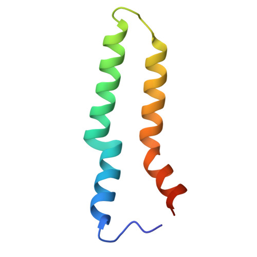
During conjugation, plasmid DNA is transferred from donor to recipient bacteria via the plasmid-encoded mating pilus, formed as thin helical assemblies of polymerised pilin subunits. In the IncHI1 R27 plasmid-encoded pilus, the TrhA pilin undergoes cyclisation (via a peptide bond between Gly1 and Asp69), essential for conjugation. Gly1 and Asp69 are exposed on the pilus surface and conserved in all TrhA pilins in the Plascad database. Substituting Asp69 with Asn, Ala, Gly, or Arg does not prevent cyclisation or pilus formation, which remains structurally indistinguishable from the wild type. Conjugation efficiency of the Asp69 substitutions across multiple recipient species correlates with side chain size, in the order Asp69Asn > Asp69Ala > Asp69Gly. However, Asp69Arg, as well as Asp69Lys and Gly1Lys substitutions abolish conjugation, likely due to the positively charged pilus surface (opposite to the wild-type negative charge) forming unfavourable electrostatic interactions with the recipient outer membrane's inner leaflet, composed solely of zwitterionic phosphatidylethanolamine (PE). Consistently, conjugation is rescued in recipients lacking PE. These findings indicate strong selective pressure to maintain Gly1 and Asp69, as efficient DNA transfer depends on precise electrostatic and steric constraints of the pilus surface.
- Department of Life Sciences, Imperial College London, London, UK.
Organizational Affiliation: