9VNQ | pdb_00009vnq

Bacillus Subtilis Ku core homodimer complexed with double strand DNA


Experimental Data Snapshot

  • Method: ELECTRON MICROSCOPY
  • Resolution: 2.74 Å
  • Aggregation State: PARTICLE 
  • Reconstruction Method: SINGLE PARTICLE 

wwPDB Validation   3D Report Full Report


This is version 1.1 of the entry. See complete history


Literature

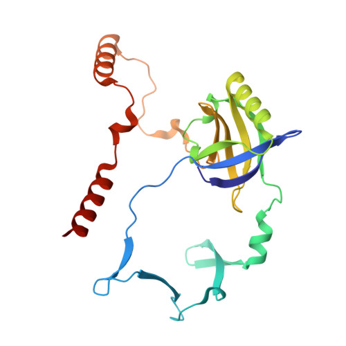
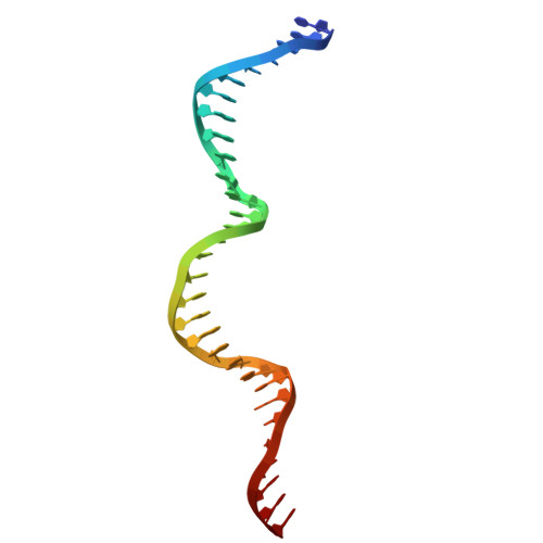
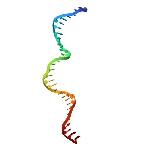
Structure of Bacillus subtilis Ku-mediated DNA synaptic complex.

Kim, W.J.Kim, J.Jo, M.Kim, Y.Kim, M.S.

(2026) Nucleic Acids Res 54

  • DOI: https://doi.org/10.1093/nar/gkaf1036
  • Primary Citation of Related Structures:  
    9VNQ

  • PubMed Abstract: 

    DNA double-strand breaks (DSBs) pose a severe threat to genomic integrity, and cells rely on two major pathways for repair: homologous recombination and non-homologous end joining (NHEJ). While eukaryotic NHEJ requires a multi-component assembly including the Ku70/80 heterodimer, bacterial NHEJ operates with a simpler toolkit comprising a Ku homodimer and the multifunctional LigD. Despite this simplicity, the mechanism by which broken DNA ends are bridged together has remained unclear in bacterial NHEJ. Here, we present a cryo-electron microscopy structure of the Bacillus subtilis Ku (bsKu)-DNA complex at 2.74 Å resolution, capturing two blunt DNA ends bridged by a Ku protein alone. Supported by further biochemical assays, we propose an integrated model in which oligomeric arrays of Ku homodimers bridge and stabilize two DNA ends, facilitating efficient DSB repair in Bacillus subtilis. This work reveals a bsKu-mediated DNA bridging mechanism distinct from the eukaryotic system and provides critical structural insight into prokaryotic DNA repair.


  • Organizational Affiliation
    • Department of Life Sciences, Pohang University of Science and Technology, Pohang, Gyeongbuk, 37673, Republic of Korea.

Macromolecules

Find similar proteins by:  (by identity cutoff)  |  3D Structure
Entity ID: 1
MoleculeChains Sequence LengthOrganismDetailsImage
Non-homologous end joining protein Ku
A, B, C, D
227Bacillus subtilisMutation(s): 0 
Gene Names: kuykoVBSU13410
UniProt
Find proteins for O34859 (Bacillus subtilis (strain 168))
Explore O34859 
Go to UniProtKB:  O34859
Entity Groups  
Sequence Clusters30% Identity50% Identity70% Identity90% Identity95% Identity100% Identity
UniProt GroupO34859
Sequence Annotations
Expand
  • Reference Sequence
Find similar nucleic acids by:  (by identity cutoff)  |  3D Structure
Entity ID: 2
MoleculeChains LengthOrganismImage
DNA (31-MER)31synthetic construct
Sequence Annotations
Expand
  • Reference Sequence
Find similar nucleic acids by:  (by identity cutoff)  |  3D Structure
Entity ID: 3
MoleculeChains LengthOrganismImage
DNA (31-MER)31synthetic construct
Sequence Annotations
Expand
  • Reference Sequence
Experimental Data & Validation

Experimental Data

  • Method: ELECTRON MICROSCOPY
  • Resolution: 2.74 Å
  • Aggregation State: PARTICLE 
  • Reconstruction Method: SINGLE PARTICLE 
EM Software:
TaskSoftware PackageVersion
RECONSTRUCTIONcryoSPARC

Structure Validation

View Full Validation Report


Entry History & Funding Information

Deposition Data


Funding OrganizationLocationGrant Number
Not funded--

Revision History  (Full details and data files)

  • Version 1.0: 2025-10-29
    Type: Initial release
  • Version 1.1: 2026-01-21
    Changes: Data collection, Database references