9VF8 | pdb_00009vf8

Structure of Meiothermus ruber Mrub_1259 LOV domain (MrLOV)


Experimental Data Snapshot

  • Method: X-RAY DIFFRACTION
  • Resolution: 3.35 Å
  • R-Value Free: 
    0.272 (Depositor), 0.266 (DCC) 
  • R-Value Work: 
    0.242 (Depositor), 0.255 (DCC) 

Starting Model: in silico
View more details

wwPDB Validation 3D Report Full Report

Validation slider image for 9VF8

Ligand Structure Quality Assessment 


This is version 1.1 of the entry. See complete history

Literature

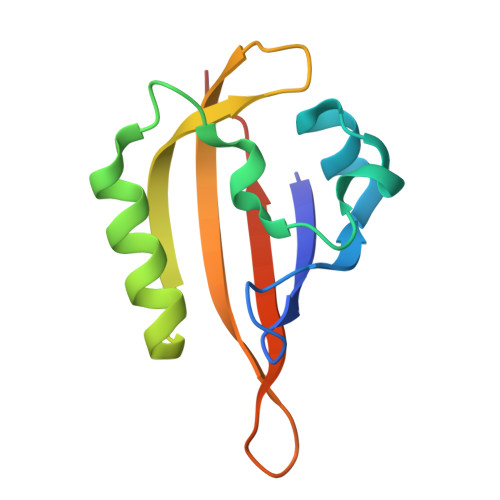
Structural characterization of Meiothermus ruber LOV domain.

Semenov, O.Nazarenko, V.Yudenko, A.Kovalev, K.Goncharov, I.Natarov, I.Mikhailov, A.Kuznetsova, E.Nikolaev, A.Yang, Y.Sluchanko, N.N.Borshchevskiy, V.Remeeva, A.Gushchin, I.

(2025) J Struct Biol 218: 108268-108268

  • DOI: https://doi.org/10.1016/j.jsb.2025.108268
  • Primary Citation Related Structures: 
    9VF7, 9VF8

  • PubMed Abstract: 

    Light Oxygen Voltage (LOV) domains are important widespread receptors of blue light that also found applications in optogenetics and imaging. While LOV domains from mesophiles are relatively well characterized, their counterparts from thermophilic microorganisms remain understudied. Here, we express two constructs of a LOV domain belonging to a histidine kinase from Meiothermus ruber, MrLOV and MrLOVe, and show that they are photoactive, with recovery time values of 21 and 27 min, respectively, and thermostable. Crystal structures reveal that MrLOV, which lacks helices A'α and Jα, forms a parallel dimer, whereas MrLOVe is a tetramer organized as an antiparallel dimer of two parallel dimers interacting via helices Jα. One MrLOVe dimer is symmetric, and the other is asymmetric, with conformational differences mirroring activation-related changes in other LOV domains. Our data provide the structural basis for understanding and engineering of thermophilic LOVs and pave the way for development of thermostable and photostable LOV-derived optogenetic tools and flavin-based fluorescent proteins.


  • Organizational Affiliation
    • Research Center for Molecular Mechanisms of Aging and Age-Related Diseases, Moscow Institute of Physics and Technology, 141700 Dolgoprudny, Russia.

Macromolecule Content 

  • Total Structure Weight: 13.17 kDa 
  • Atom Count: 860 
  • Modeled Residue Count: 105 
  • Deposited Residue Count: 112 
  • Unique protein chains: 1

Macromolecules

Find similar proteins by:|  3D Structure
Entity ID: 1
MoleculeChains  Sequence LengthOrganismDetailsImage
histidine kinase112Meiothermus ruber DSM 1279Mutation(s): 0 
Gene Names: Mrub_1259K649_05955
EC: 2.7.13.3
UniProt
Find proteins for D3PRD8 (Meiothermus ruber (strain ATCC 35948 / DSM 1279 / VKM B-1258 / 21))
Explore D3PRD8 
Go to UniProtKB:  D3PRD8
Entity Groups
Sequence Clusters30% Identity50% Identity70% Identity90% Identity95% Identity100% Identity
UniProt GroupD3PRD8
Sequence Annotations
Expand
Reference Sequence

Small Molecules

Ligands 1 Unique
IDChains Name / Formula / InChI Key2D Diagram3D Interactions
FMN
(Subject of Investigation/LOI)

Query on FMN


Download:Ideal Coordinates CCD File
B [auth A]FLAVIN MONONUCLEOTIDE
C17 H21 N4 O9 P
FVTCRASFADXXNN-SCRDCRAPSA-N

Experimental Data & Validation

Experimental Data

  • Method: X-RAY DIFFRACTION
  • Resolution: 3.35 Å
  • R-Value Free:  0.272 (Depositor), 0.266 (DCC) 
  • R-Value Work:  0.242 (Depositor), 0.255 (DCC) 
Space Group: P 43 21 2
Unit Cell:
Length ( Å )Angle ( ˚ )
a = 78.593α = 90
b = 78.593β = 90
c = 73.381γ = 90
Software Package:
Software NamePurpose
XDSdata reduction
STARANISOdata scaling
Aimlessdata scaling
REFMACrefinement
MOLREPphasing

Structure Validation

View Full Validation Report


Ligand Structure Quality Assessment 


Entry History 

& Funding Information

Deposition Data


Funding OrganizationLocationGrant Number
Not funded--

Revision History  (Full details and data files)

  • Version 1.0: 2025-06-25
    Type: Initial release
  • Version 1.1: 2025-12-17
    Changes: Database references