9VBH | pdb_00009vbh

Cryo-EM structure of human PLD3 bound to ssDNA (poly(T))


Experimental Data Snapshot

  • Method: ELECTRON MICROSCOPY
  • Resolution: 2.85 Å
  • Aggregation State: PARTICLE 
  • Reconstruction Method: SINGLE PARTICLE 

wwPDB Validation   3D Report Full Report


This is version 1.0 of the entry. See complete history


Literature

Mechanistic insights into single-stranded DNA degradation by lysosomal exonucleases PLD3 and PLD4 from structural snapshots.

Hirano, Y.Ezaki, W.Sato, R.Ohto, U.Miyake, K.Shimizu, T.

(2025) Nat Commun 

  • DOI: https://doi.org/10.1038/s41467-025-66261-2
  • Primary Citation of Related Structures:  
    9VBG, 9VBH, 9VBI, 9VBJ, 9VBK

  • PubMed Abstract: 

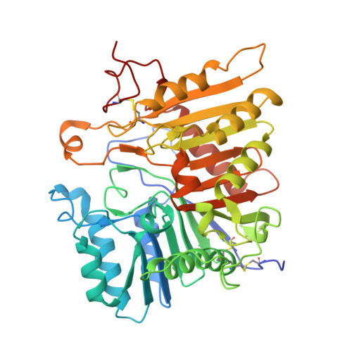
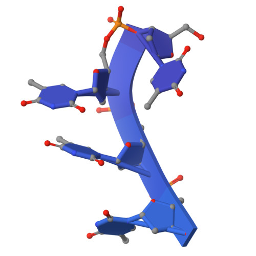
    Lysosomal exonuclease phospholipase D (PLD) family PLD3 and PLD4 degrade single-stranded RNA or DNA and regulate TLR7 or TLR9 responses. Polymorphisms of these enzymes are associated with human diseases: PLD4 is associated with inflammatory diseases, and PLD3 is associated with neurodegenerative diseases. Here, we determine the structures of substrate-bound PLD3 and PLD4 by cryo-electron microscopy. Our structures reveal that PLD3 rebuilds a substrate-binding pocket, depending on the substrate, mainly via motion of the Phe335-containing loop. Furthermore, we captured the structure in a metastable state that appears during substrate rearrangement following product release. Together, our findings identify the residues that underlie the distinct activities of PLD3 and PLD4. This study provides a mechanistic basis for the exonuclease activity of PLD3 and PLD4 in single-stranded DNA degradation.


  • Organizational Affiliation
    • Graduate School of Pharmaceutical Sciences, The University of Tokyo, Tokyo, Japan.

Macromolecules

Find similar proteins by:  (by identity cutoff)  |  3D Structure
Entity ID: 1
MoleculeChains Sequence LengthOrganismDetailsImage
5'-3' exonuclease PLD3
A, C
436Homo sapiensMutation(s): 2 
Gene Names: PLD3
EC: 3.1.16.1 (PDB Primary Data), 3.1.4 (PDB Primary Data)
UniProt & NIH Common Fund Data Resources
Find proteins for Q8IV08 (Homo sapiens)
Explore Q8IV08 
Go to UniProtKB:  Q8IV08
PHAROS:  Q8IV08
GTEx:  ENSG00000105223 
Entity Groups  
Sequence Clusters30% Identity50% Identity70% Identity90% Identity95% Identity100% Identity
UniProt GroupQ8IV08
Glycosylation
Glycosylation Sites: 4Go to GlyGen: Q8IV08-1
Sequence Annotations
Expand
  • Reference Sequence
Find similar nucleic acids by:  (by identity cutoff)  |  3D Structure
Entity ID: 2
MoleculeChains LengthOrganismImage
DNA (5'-D(*TP*TP*TP*T)-3')
B, D
49synthetic construct
Sequence Annotations
Expand
  • Reference Sequence
Oligosaccharides

Help

Entity ID: 3
MoleculeChains Length2D Diagram Glycosylation3D Interactions
2-acetamido-2-deoxy-beta-D-glucopyranose-(1-4)-2-acetamido-2-deoxy-beta-D-glucopyranose
E, H, I, L
2N-Glycosylation
Entity ID: 4
MoleculeChains Length2D Diagram Glycosylation3D Interactions
beta-D-mannopyranose-(1-4)-2-acetamido-2-deoxy-beta-D-glucopyranose-(1-4)-2-acetamido-2-deoxy-beta-D-glucopyranose
F, J
3N-Glycosylation
Entity ID: 5
MoleculeChains Length2D Diagram Glycosylation3D Interactions
alpha-D-mannopyranose-(1-3)-[alpha-D-mannopyranose-(1-6)]beta-D-mannopyranose-(1-4)-2-acetamido-2-deoxy-beta-D-glucopyranose-(1-4)-2-acetamido-2-deoxy-beta-D-glucopyranose
G, K
5N-Glycosylation
Experimental Data & Validation

Experimental Data

  • Method: ELECTRON MICROSCOPY
  • Resolution: 2.85 Å
  • Aggregation State: PARTICLE 
  • Reconstruction Method: SINGLE PARTICLE 
EM Software:
TaskSoftware PackageVersion
RECONSTRUCTIONcryoSPARC

Structure Validation

View Full Validation Report


Entry History & Funding Information

Deposition Data


Funding OrganizationLocationGrant Number
Japan Society for the Promotion of Science (JSPS)Japan22K06110
Japan Society for the Promotion of Science (JSPS)Japan25K09523
Japan Science and TechnologyJapanJPMJCR21E4

Revision History  (Full details and data files)

  • Version 1.0: 2025-12-24
    Type: Initial release