Structural insights into C-terminus-mediated RNA target cleavage by a mesophilic prokaryotic argonaute.
Chen, T., Tao, X., Li, S., Duanmu, Q., Wang, J., Chen, K., Mu, K., Yang, H., Li, Y., Liu, Y., Ma, L., Wu, S.(2026) Nat Commun 17
- PubMed: 41932907 Search on PubMed
- DOI: https://doi.org/10.1038/s41467-026-71422-y
- Primary Citation Related Structures:
9V65, 9V66, 9V7S, 9V7Z, 9V8A, 9V93, 9V9G, 9V9I - PubMed Abstract:
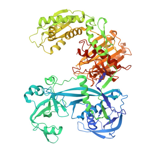
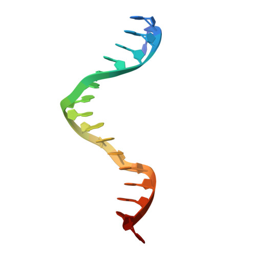
Prokaryotic Argonaute proteins (pAgos) are programmable nucleases that always utilize DNA guides to cleave DNA targets. Recent studies show that some pAgos preferentially utilize DNA guides to cleave RNA targets rather than DNA targets. VbAgo, derived from a Verrucomicrobia bacterium, is a nuclease capable of specifically cleaving single-stranded RNA and highly structured RNA substrates at 37 °C, making it an ideal candidate for developing RNA manipulation toolkits. An in-depth investigation of its mechanism contributes to understanding the functional characteristics of gDtR-type Ago proteins. Here, we present cryo-electron microscopy structures of VbAgo, the VbAgo-guide DNA binary complex, multiple wild-type VbAgo-guide DNA-target RNA ternary complexes, and the catalytically inactive mutant (VbAgo-DM) guide DNA-target RNA ternary complex, with resolutions ranging from 2.5 to 3.2 Å. By integrating these cryo-EM structures with biochemical data, we elucidate the entire catalytic process of VbAgo, revealing its unique C-terminal regulatory mechanism. Specifically, in its apo state, VbAgo's C-terminus occupies the nucleic acid binding channel, partially impeding its catalytic activity while enhancing its stability. The binding of guide DNA displaces the C-terminus, and subsequent binding of target RNA, along with conformational changes in the N-terminal and PAZ domains, facilitates VbAgo dimerization. Following this, the C-terminus transitions from a loop to a helix, enabling maturation of the catalytic center and inducing movements in the MID-PIWI' interactions at the dimer interfaces, ultimately leading to dimer dissociation. Concurrently, cleavage of the target RNA and subsequent product release occur, after which VbAgo reverts to its binary state to initiate the next cleavage cycle. Moreover, we demonstrate that VbAgo exhibits guide DNA mediated RNA knockdown activity in mammalian cells. In summary, our study provides a comprehensive understanding of the molecular mechanisms governing self-inhibition, guide binding, target recognition, and product release in VbAgo. These findings offer valuable insights into the diverse mechanisms of pAgos, broadening their functional scope and enhancing the biotechnological potential of pAgo proteins.
- Hubei Key Laboratory of Industrial Biotechnology, School of Life Sciences, Hubei University, Wuhan, Hubei, China.
Organizational Affiliation: