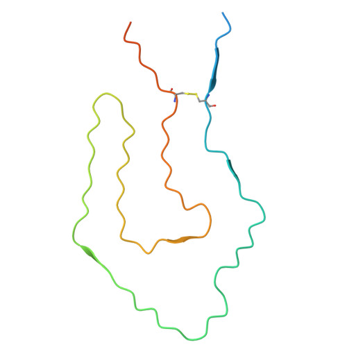
9V91 | pdb_00009v91

Cryo-EM structure of renal amyloid fibril from an immunoglobulin light chain amyloidosis patient in polymorph B


Experimental Data Snapshot

  • Method: ELECTRON MICROSCOPY
  • Resolution: 3.00 Å
  • Aggregation State: HELICAL ARRAY 
  • Reconstruction Method: HELICAL 

wwPDB Validation 3D Report Full Report

Validation slider image for 9V91

This is version 1.0 of the entry. See complete history

Literature

Cryo-EM structure of renal amyloid fibril from an immunoglobulin light chain amyloidosis patient in polymorph B

Zhao, K.Yu, C.Y.Ma, Y.Y.Huang, H.

To be published.

Macromolecule Content 

  • Total Structure Weight: 68.19 kDa 
  • Atom Count: 3,588 
  • Modeled Residue Count: 480 
  • Deposited Residue Count: 660 
  • Unique protein chains: 1

Macromolecules

Find similar proteins by:|  3D Structure
Entity ID: 1
MoleculeChains  Sequence LengthOrganismDetailsImage
IG c956_light_IGLV1-44_IGLJ3
A, B, C, D, E
A, B, C, D, E, F
110Homo sapiensMutation(s): 0 
UniProt
Find proteins for A0A5C2GJC2 (Homo sapiens)
Explore A0A5C2GJC2 
Go to UniProtKB:  A0A5C2GJC2
Entity Groups
Sequence Clusters30% Identity50% Identity70% Identity90% Identity95% Identity100% Identity
UniProt GroupA0A5C2GJC2
Sequence Annotations
Expand
Reference Sequence

Experimental Data & Validation

Experimental Data

  • Method: ELECTRON MICROSCOPY
  • Resolution: 3.00 Å
  • Aggregation State: HELICAL ARRAY 
  • Reconstruction Method: HELICAL 
EM Software:
TaskSoftware PackageVersion
MODEL REFINEMENTPHENIX1.20.1_4487

Structure Validation

View Full Validation Report


Entry History 

& Funding Information

Deposition Data


Funding OrganizationLocationGrant Number
Not funded--

Revision History  (Full details and data files)

  • Version 1.0: 2025-12-24
    Type: Initial release