9V7V | pdb_00009v7v

Structure of FtsZ1 in complex with GMPCPP from Candidatus Odinarchaeota


Experimental Data Snapshot

  • Method: ELECTRON MICROSCOPY
  • Resolution: 3.60 Å
  • Aggregation State: FILAMENT 
  • Reconstruction Method: HELICAL 

Starting Model: in silico
View more details

wwPDB Validation 3D Report Full Report

Validation slider image for 9V7V

This is version 1.2 of the entry. See complete history

Literature

Distinct filament morphology and membrane tethering features of the dual FtsZ paralogs in Odinarchaeota.

Kumari, J.Uthaman, A.Bose, S.Kundu, A.Sharma, V.Dutta, S.Dhar, A.Roy, S.Srinivasan, R.Pande, S.Vinothkumar, K.R.Gayathri, P.Palani, S.

(2025) EMBO J 44: 5940-5964

  • DOI: https://doi.org/10.1038/s44318-025-00529-7
  • Primary Citation Related Structures: 
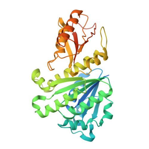
    9V7V

  • PubMed Abstract: 

    The Asgard phylum has emerged as a model to study eukaryogenesis because of their close relatedness with the eukaryotes. In this study, we use FtsZ proteins from a member of the class Odinarchaeia as representatives to investigate the probable origin, evolution, and assembly of the FtsZ/tubulin protein superfamily in Asgard archaea. We performed a comparative analysis of the biochemical properties and cytoskeletal assembly of FtsZ1 and FtsZ2, the two FtsZ isoforms in the Odinarchaeota metagenome. Our electron microscopy analysis reveals that OdinFtsZ1 assembles into curved single protofilaments, while OdinFtsZ2 forms stacked spiral ring-like structures. Upon sequence analysis, we identified an N-terminal amphipathic helix in OdinFtsZ1, which mediates direct membrane tethering. In contrast, OdinFtsZ2 is recruited to the membrane by the anchor OdinSepF via OdinFtsZ2's C-terminal tail. Overall, we report the presence of two distant evolutionary paralogs of FtsZ in Odinarchaeota, with distinct filament assemblies and differing modes of membrane targeting. Our findings highlight the diversity of FtsZ proteins in the archaeal phylum Asgardarchaeota, providing valuable insights into the evolution and differentiation of tubulin-family proteins.


  • Organizational Affiliation
    • Department of Biochemistry, Indian Institute of Science, Bengaluru, Karnataka, 560012, India.

Macromolecule Content 

  • Total Structure Weight: 157.06 kDa 
  • Atom Count: 8,912 
  • Modeled Residue Count: 1,196 
  • Deposited Residue Count: 1,452 
  • Unique protein chains: 1

Macromolecules

Find similar proteins by:|  3D Structure
Entity ID: 1
MoleculeChains  Sequence LengthOrganismDetailsImage
Cell division protein FtsZ
A, B, C, D
363Candidatus OdinarchaeotaMutation(s): 0 
Gene Names: ftsZOdinLCB4_002530
UniProt
Find proteins for Q8TX55 (Methanopyrus kandleri (strain AV19 / DSM 6324 / JCM 9639 / NBRC 100938))
Explore Q8TX55 
Go to UniProtKB:  Q8TX55
Entity Groups
Sequence Clusters30% Identity50% Identity70% Identity90% Identity95% Identity100% Identity
UniProt GroupQ8TX55
Sequence Annotations
Expand
Reference Sequence

Experimental Data & Validation

Experimental Data

  • Method: ELECTRON MICROSCOPY
  • Resolution: 3.60 Å
  • Aggregation State: FILAMENT 
  • Reconstruction Method: HELICAL 
EM Software:
TaskSoftware PackageVersion
RECONSTRUCTIONcryoSPARC4.5.3
MODEL REFINEMENTPHENIX1.21.2_5419

Structure Validation

View Full Validation Report


Entry History 

& Funding Information

Deposition Data


Funding OrganizationLocationGrant Number
Department of Biotechnology (DBT, India)IndiaDBT/PR12422/MED/31/287/2014

Revision History  (Full details and data files)

  • Version 1.0: 2025-07-30
    Type: Initial release
  • Version 1.1: 2025-08-20
    Changes: Data collection, Database references
  • Version 1.2: 2025-11-19
    Changes: Data collection, Database references