9UZA | pdb_00009uza

Crystal structure of Crimean-Congo hemorrhagic fever virus cap-snatching endonuclease


Experimental Data Snapshot

  • Method: X-RAY DIFFRACTION
  • Resolution: 3.05 Å
  • R-Value Free: 
    0.295 (Depositor), 0.289 (DCC) 
  • R-Value Work: 
    0.226 (Depositor), 0.223 (DCC) 
  • R-Value Observed: 
    0.229 (Depositor) 

Starting Model: in silico
View more details

wwPDB Validation   3D Report Full Report


This is version 1.1 of the entry. See complete history


Literature

Structure and function of the nairovirus cap-snatching endonuclease.

Kuang, W.Tian, Z.Zhang, G.Wu, F.Li, J.Tang, J.Zhang, H.Zhuo, X.Hu, Z.Wang, M.Zhao, H.Deng, Z.

(2026) Nucleic Acids Res 54

  • DOI: https://doi.org/10.1093/nar/gkaf1515
  • Primary Citation Related Structures: 
    9UZA, 9UZB, 9UZC, 9UZD, 9UZE, 9UZF, 9UZG, 9UZH, 9UZI

  • PubMed Abstract: 

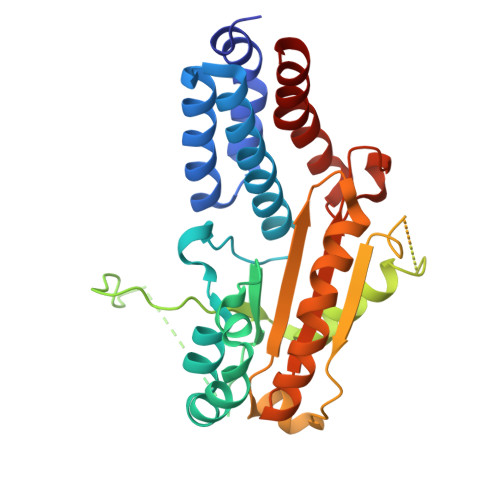
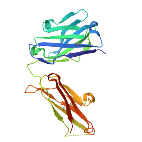
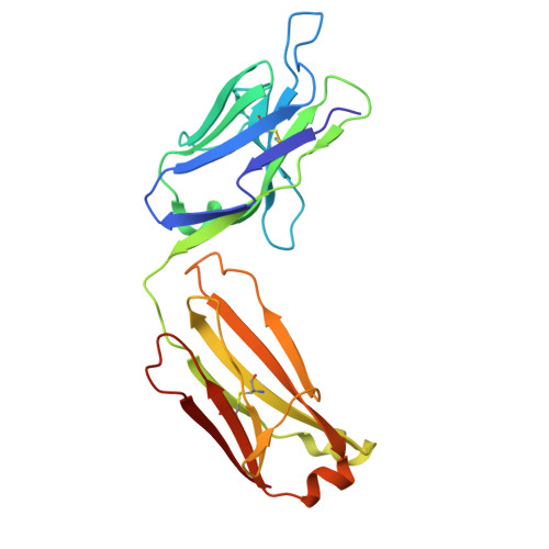
    Nairoviruses include several human pathogens such as Crimean-Congo hemorrhagic fever virus (CCHFV) and Kasokero virus (KASV). The cap-snatching endonuclease (EN) domain of the viral polymerase is essential for transcription and represents a promising antiviral target. However, the structural and functional mechanisms of nairovirus ENs remain poorly understood. Here, we describe biochemical and structural studies of the ENs from CCHFV and KASV. Biochemical assays demonstrate that the RNA endonuclease activity of both ENs is activated by manganese ions and exhibits a preference for uridine-rich RNA substrates. This activity is inhibited by three metal-chelating inhibitors (DPBA, L-742,001, and BXA), with BXA displaying the highest binding affinity and inhibitory potency. We further determine nine crystal structures of CCHFV and KASV ENs in apo, metal ion-bound, and inhibitor-bound states. Comparative structural analysis uncovers a two-metal-ion binding mode unique to nairovirus ENs, in which conserved residues coordinate two manganese ions via bridging water molecules. In the inhibitor-bound structures of KASV EN, BXA forms additional stabilizing interactions with the enzyme, explaining its superior inhibitory effect. Functional assays further confirm that the two-metal-ion mechanism is critical for viral transcription. These findings provide a structural foundation for the rational design of antivirals against CCHFV and related pathogens.


  • Organizational Affiliation
    • State Key Laboratory of Virology and Biosafety, Wuhan Institute of Virology, Chinese Academy of Sciences, Wuhan 430207, Hubei, China.

Macromolecules
Find similar proteins by:  (by identity cutoff)  |  3D Structure
Entity ID: 1
MoleculeChains Sequence LengthOrganismDetailsImage
RNA-directed RNA polymerase LA,
D [auth B]
319Orthonairovirus haemorrhagiaeMutation(s): 0 
EC: 2.7.7.48 (PDB Primary Data), 3.4.19.12 (PDB Primary Data)
UniProt
Find proteins for D5MEH7 (Orthonairovirus haemorrhagiae)
Explore D5MEH7 
Go to UniProtKB:  D5MEH7
Entity Groups  
Sequence Clusters30% Identity50% Identity70% Identity90% Identity95% Identity100% Identity
UniProt GroupD5MEH7
Sequence Annotations
Expand
  • Reference Sequence
Find similar proteins by:  (by identity cutoff)  |  3D Structure
Entity ID: 2
MoleculeChains Sequence LengthOrganismDetailsImage
mAb G5 Fab heavy chainB [auth H],
E [auth C]
228Mus musculusMutation(s): 0 
Entity Groups  
Sequence Clusters30% Identity50% Identity70% Identity90% Identity95% Identity100% Identity
Sequence Annotations
Expand
  • Reference Sequence
Find similar proteins by:  (by identity cutoff)  |  3D Structure
Entity ID: 3
MoleculeChains Sequence LengthOrganismDetailsImage
mAb G5 Fab light chainC [auth L],
F [auth D]
219Mus musculusMutation(s): 0 
Entity Groups  
Sequence Clusters30% Identity50% Identity70% Identity90% Identity95% Identity100% Identity
Sequence Annotations
Expand
  • Reference Sequence
Experimental Data & Validation

Experimental Data

  • Method: X-RAY DIFFRACTION
  • Resolution: 3.05 Å
  • R-Value Free:  0.295 (Depositor), 0.289 (DCC) 
  • R-Value Work:  0.226 (Depositor), 0.223 (DCC) 
  • R-Value Observed: 0.229 (Depositor) 
Space Group: P 21 21 21
Unit Cell:
Length ( Å )Angle ( ˚ )
a = 98.767α = 90
b = 99.779β = 90
c = 180.419γ = 90
Software Package:
Software NamePurpose
PHENIXrefinement
PDB_EXTRACTdata extraction
XDSdata reduction
XDSdata scaling
PHASERphasing

Structure Validation

View Full Validation Report


Entry History & Funding Information

Deposition Data


Funding OrganizationLocationGrant Number
Not funded--

Revision History  (Full details and data files)

  • Version 1.0: 2025-12-31
    Type: Initial release
  • Version 1.1: 2026-04-01
    Changes: Database references