9UYR | pdb_00009uyr

In situ structure of egg-white lysozyme using a goniometer-compatible chip-based platform

  • Classification: HYDROLASE
  • Organism(s): Gallus gallus
  • Mutation(s): No 

  • Deposited: 2025-05-15 Released: 2026-03-25 
  • Deposition Author(s): Ghosh, S.
  • Funding Organization(s): European Research Council (ERC)

Experimental Data Snapshot

  • Method: X-RAY DIFFRACTION
  • Resolution: 1.59 Å
  • R-Value Free: 
    0.194 (Depositor), 0.192 (DCC) 
  • R-Value Work: 
    0.169 (Depositor), 0.170 (DCC) 
  • R-Value Observed: 
    0.170 (Depositor) 

Starting Model: experimental
View more details

wwPDB Validation 3D Report Full Report

Validation slider image for 9UYR

This is version 1.1 of the entry. See complete history

Literature

A user-friendly goniometer-compatible fixed-target platform for macromolecular crystallography at synchrotrons.

Ghosh, S.Banacore, A.Norder, P.Bjelcic, M.Kabbinale, A.Nileshwar, P.Wehlander, G.de Sanctis, D.Basu, S.Orlans, J.Vallejos, A.Chavas, L.M.G.Neutze, R.Branden, G.

(2026) J Appl Crystallogr 59: 303-315

  • DOI: https://doi.org/10.1107/S1600576725011513
  • Primary Citation Related Structures: 
    9TBL, 9UYR, 9VDJ, 9VDX

  • PubMed Abstract: 

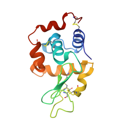
    Fixed-target platforms provide convenient support for microcrystals during serial X-ray crystallography studies using synchrotron radiation. Here, we describe a simple user-friendly 3D-printed support where the crystals are sandwiched between two layers of thin X-ray-transparent membrane resulting in very low scattering background. The platform is compatible with magnetic mounting onto the standard goniometer of macromolecular crystallography beamlines. Our design utilizes a 96-well frame that facilitates hanging-drop experiments directly on the membrane using conventional crystallization plates, thereby eliminating multiple pipetting and crystal handling steps. Crystals can be enclosed in a sandwich and packed into 'cassettes', preventing the risk of the sample drying out during room-temperature transportation to synchrotron sources. The versatility of the platform is demonstrated by five structures solved using different crystallization and data-collection strategies. Lysozyme single-crystal rotational crystallography at room temperature is shown, as well as microcrystal serial data collection under cryogenic conditions. On-chip microcrystallization is illustrated by use of a photosynthetic reaction center as an example. Finally, serial crystallography data collection at room temperature from microcrystals of the membrane protein cytochrome c oxidase crystallized in lipidic cubic phase is presented.


  • Organizational Affiliation
    • Department of Chemistry and Molecular Biology Gothenburg University Sweden.

Macromolecule Content 

  • Total Structure Weight: 14.39 kDa 
  • Atom Count: 1,059 
  • Modeled Residue Count: 129 
  • Deposited Residue Count: 129 
  • Unique protein chains: 1

Macromolecules

Find similar proteins by:|  3D Structure
Entity ID: 1
MoleculeChains  Sequence LengthOrganismDetailsImage
Lysozyme C129Gallus gallusMutation(s): 0 
EC: 3.2.1.17
UniProt
Find proteins for P00698 (Gallus gallus)
Explore P00698 
Go to UniProtKB:  P00698
Entity Groups
Sequence Clusters30% Identity50% Identity70% Identity90% Identity95% Identity100% Identity
UniProt GroupP00698
Sequence Annotations
Expand
Reference Sequence

Experimental Data & Validation

Experimental Data

  • Method: X-RAY DIFFRACTION
  • Resolution: 1.59 Å
  • R-Value Free:  0.194 (Depositor), 0.192 (DCC) 
  • R-Value Work:  0.169 (Depositor), 0.170 (DCC) 
  • R-Value Observed: 0.170 (Depositor) 
Space Group: P 43 21 2
Unit Cell:
Length ( Å )Angle ( ˚ )
a = 79.116α = 90
b = 79.116β = 90
c = 37.925γ = 90
Software Package:
Software NamePurpose
MxCuBEdata collection
autoPROCdata processing
PHENIXphasing
PHENIXrefinement
Cootmodel building

Structure Validation

View Full Validation Report


Entry History 

& Funding Information

Deposition Data

  • Released Date: 2026-03-25 
  • Deposition Author(s): Ghosh, S.

Funding OrganizationLocationGrant Number
European Research Council (ERC)European Union--

Revision History  (Full details and data files)

  • Version 1.0: 2026-03-25
    Type: Initial release
  • Version 1.1: 2026-04-22
    Changes: Database references