9UWI | pdb_00009uwi

Cryo-EM structure of human V1aR bound with atosiban at a resolution of 2.8 angstrom


Experimental Data Snapshot

  • Method: ELECTRON MICROSCOPY
  • Resolution: 2.80 Å
  • Aggregation State: PARTICLE 
  • Reconstruction Method: SINGLE PARTICLE 

wwPDB Validation   3D Report Full Report


This is version 1.0 of the entry. See complete history


Literature

Molecular basis of antagonism of the dimeric human arginine vasopressin receptor 1A.

Zhong, P.Chu, B.Yu, Z.Qiao, Y.Ding, Y.Zhang, Y.Wu, X.

(2026) Nat Commun 

  • DOI: https://doi.org/10.1038/s41467-026-68331-5
  • Primary Citation of Related Structures:  
    9UWI

  • PubMed Abstract: 

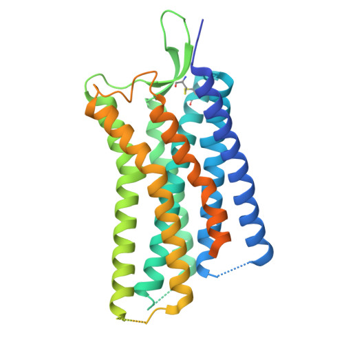
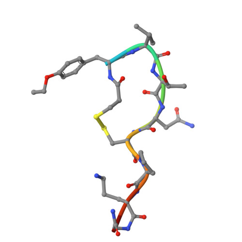
    Arginine vasopressin (AVP) and oxytocin (OT) are peptide hormones critical for various physiological processes. Vasopressin receptor 1 A (V1aR), a primary AVP target, is promising for central nervous system (CNS) disorders therapies, yet the mechanisms of antagonism and oligomerization remain poorly understood. Here, we present structures of human V1aR in its apo state and in complexes with antagonists: atosiban, balovaptan, and SRX246. Structural analyses reveal a dimeric V1aR assembly, validated by functional assays and imaging in cells. The apo structure shows a flat extracellular loop 2 (ECL2) with unpaired cysteines, undergoing significant conformational changes upon ligand binding. Antagonist-bound structures, combined with mutagenesis and radioligand binding assays, uncover distinct binding modes and key determinants for antagonism and selectivity. These findings provide a comprehensive understanding of V1aR assembly and dynamic regulation, offering valuable insights for structure-guided development of new antagonists targeting dimeric V1aR for CNS disorders.


  • Organizational Affiliation
    • Fudan University, Shanghai, China.

Macromolecules
Find similar proteins by:  (by identity cutoff)  |  3D Structure
Entity ID: 1
MoleculeChains Sequence LengthOrganismDetailsImage
Vasopressin V1a receptor
A, B
386Homo sapiensMutation(s): 2 
Gene Names: AVPR1AAVPR1
UniProt & NIH Common Fund Data Resources
Find proteins for P37288 (Homo sapiens)
Explore P37288 
Go to UniProtKB:  P37288
PHAROS:  P37288
GTEx:  ENSG00000166148 
Entity Groups  
Sequence Clusters30% Identity50% Identity70% Identity90% Identity95% Identity100% Identity
UniProt GroupP37288
Sequence Annotations
Expand
  • Reference Sequence

Find similar proteins by:  Sequence   |   3D Structure  

Entity ID: 2
MoleculeChains Sequence LengthOrganismDetailsImage
Atosiban
C, D
10synthetic constructMutation(s): 0 
Entity Groups  
Sequence Clusters30% Identity50% Identity70% Identity90% Identity95% Identity100% Identity
Sequence Annotations
Expand
  • Reference Sequence
Experimental Data & Validation

Experimental Data

  • Method: ELECTRON MICROSCOPY
  • Resolution: 2.80 Å
  • Aggregation State: PARTICLE 
  • Reconstruction Method: SINGLE PARTICLE 
EM Software:
TaskSoftware PackageVersion
MODEL REFINEMENTPHENIX1.20.1_4487

Structure Validation

View Full Validation Report


Entry History & Funding Information

Deposition Data


Funding OrganizationLocationGrant Number
National Science Foundation (NSF, China)China32471303

Revision History  (Full details and data files)

  • Version 1.0: 2026-01-28
    Type: Initial release