9USU | pdb_00009usu

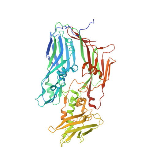
Two interacting D13 trimers at mode I interface in Twister assembly


Experimental Data Snapshot

  • Method: ELECTRON MICROSCOPY
  • Resolution: 2.90 Å
  • Aggregation State: PARTICLE 
  • Reconstruction Method: SINGLE PARTICLE 

wwPDB Validation 3D Report Full Report

Validation slider image for 9USU

This is version 1.0 of the entry. See complete history

Literature

Structures of in vitro assembly products of poxvirus scaffolding protein reveal transition from pre-assembly state to fully assembled scaffold

Jang, Y.T.Kim, S.M.Lee, S.N.Ryu, B.H.Jeong, H.S.Kang, E.S.Sul, J.H.Kim, Y.H.Jo, D.G.Hyun, J.K.

To be published.

Macromolecule Content 

  • Total Structure Weight: 372.12 kDa 
  • Atom Count: 25,296 
  • Modeled Residue Count: 3,180 
  • Deposited Residue Count: 3,312 
  • Unique protein chains: 1

Macromolecules

Find similar proteins by:|  3D Structure
Entity ID: 1
MoleculeChains  Sequence LengthOrganismDetailsImage
Scaffold protein OPG125
A, B, C, D, E
A, B, C, D, E, F
552Orthopoxvirus vacciniaMutation(s): 0 
Gene Names: OPG125MVA110LACAM3000_MVA_110
UniProt
Find proteins for Q76ZR4 (Vaccinia virus (strain Ankara))
Explore Q76ZR4 
Go to UniProtKB:  Q76ZR4
Entity Groups
Sequence Clusters30% Identity50% Identity70% Identity90% Identity95% Identity100% Identity
UniProt GroupQ76ZR4
Sequence Annotations
Expand
Reference Sequence

Experimental Data & Validation

Experimental Data

  • Method: ELECTRON MICROSCOPY
  • Resolution: 2.90 Å
  • Aggregation State: PARTICLE 
  • Reconstruction Method: SINGLE PARTICLE 
EM Software:
TaskSoftware PackageVersion
MODEL REFINEMENTPHENIX1.20.1_4487

Structure Validation

View Full Validation Report


Entry History 

& Funding Information

Deposition Data


Funding OrganizationLocationGrant Number
National Research Foundation (NRF, Korea)Korea, Republic Of--

Revision History  (Full details and data files)

  • Version 1.0: 2026-08-05
    Type: Initial release