9UJJ | pdb_00009ujj

Crystal structure of the BTB domain mouse Keap1 in complex with CDDO-Me


Experimental Data Snapshot

  • Method: X-RAY DIFFRACTION
  • Resolution: 3.43 Å
  • R-Value Free: 
    0.320 (Depositor), 0.317 (DCC) 
  • R-Value Work: 
    0.252 (Depositor), 0.253 (DCC) 
  • R-Value Observed: 
    0.255 (Depositor) 

Starting Model: experimental
View more details

wwPDB Validation 3D Report Full Report

Validation slider image for 9UJJ

Ligand Structure Quality Assessment 


This is version 1.1 of the entry. See complete history

Literature

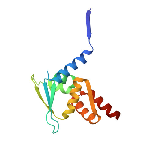
Bidirectional regulation of KEAP1 BTB domain-based sensor activity.

Suzuki, T.Takagi, K.Iso, T.Wen, H.Zhang, A.Hatakeyama, T.Oshima, H.Mizushima, T.Yamamoto, M.

(2025) Redox Biol 87: 103885-103885

  • DOI: https://doi.org/10.1016/j.redox.2025.103885
  • Primary Citation Related Structures: 
    9UJG, 9UJI, 9UJJ

  • PubMed Abstract: 

    The KEAP1-CUL3 ubiquitin ligase regulates protein stability of transcriptional factor NRF2 and plays critical roles in cellular stress response. The BTB domain of KEAP1 functions as a sensor for electrophilic chemicals. However, the precise mechanisms by which electrophiles are recognized and inhibit BTB activity remain unclear. Here, we show that electrophilic modification alters the spatial arrangement of the BTB homodimer, regulating its ligase activity. Co-crystal structural analyses and functional studies using potent NRF2-inducing CDDO-derivatives, synthetic electrophilic compounds structurally related to clinically approved molecules such as Omaveloxolone, revealed that the key sensor residue, Cys151, resides in a structurally elaborate environment within the BTB domain. Modification of Cys151 by NRF2 inducers changes the spatial configuration of the CUL3-binding sites in the BTB homodimer, reducing KEAP1-CUL3 complex affinity. In contrast, a Cys151-targeting NRF2 inhibitor induces an opposite rearrangement of the BTB homodimer. This study elucidates the molecular mechanism by which the BTB domain finely regulates KEAP1-CUL3 ubiquitin ligase activity.


  • Organizational Affiliation
    • Department of Biochemistry & Molecular Biology, Tohoku Medical Megabank Organization, Tohoku University, 2-1 Seiryo-machi, Aoba-ku, Sendai, 980-8573, Japan; Advanced Research Center for Innovations in Next-Generation Medicine (INGEM), Tohoku University, 2-1 Seiryo-machi, Aoba-ku, Sendai, 980-8573, Japan. Electronic address: takafumi.suzuki.d5@tohoku.ac.jp.

Macromolecule Content 

  • Total Structure Weight: 119.26 kDa 
  • Atom Count: 7,812 
  • Modeled Residue Count: 976 
  • Deposited Residue Count: 1,040 
  • Unique protein chains: 1

Macromolecules

Find similar proteins by:|  3D Structure
Entity ID: 1
MoleculeChains  Sequence LengthOrganismDetailsImage
Kelch-like ECH-associated protein 1
A, B, C, D, E
A, B, C, D, E, F, G, H
130Mus musculusMutation(s): 1 
Gene Names: Keap1Inrf2Kiaa0132
UniProt
Find proteins for Q9Z2X8 (Mus musculus)
Explore Q9Z2X8 
Go to UniProtKB:  Q9Z2X8
Entity Groups
Sequence Clusters30% Identity50% Identity70% Identity90% Identity95% Identity100% Identity
UniProt GroupQ9Z2X8
Sequence Annotations
Expand
Reference Sequence

Experimental Data & Validation

Experimental Data

  • Method: X-RAY DIFFRACTION
  • Resolution: 3.43 Å
  • R-Value Free:  0.320 (Depositor), 0.317 (DCC) 
  • R-Value Work:  0.252 (Depositor), 0.253 (DCC) 
  • R-Value Observed: 0.255 (Depositor) 
Space Group: C 1 2 1
Unit Cell:
Length ( Å )Angle ( ˚ )
a = 324.477α = 90
b = 72.101β = 103.294
c = 92.679γ = 90
Software Package:
Software NamePurpose
PHENIXrefinement
XDSdata reduction
XDSdata scaling
PHASERphasing

Structure Validation

View Full Validation Report


Ligand Structure Quality Assessment 


Entry History 

& Funding Information

Deposition Data


Funding OrganizationLocationGrant Number
Ministry of Education, Culture, Sports, Science and Technology (Japan)Japan21H05270
Japan Society for the Promotion of Science (JSPS)Japan22K06876
Japan Society for the Promotion of Science (JSPS)Japan25K02449
Japan Society for the Promotion of Science (JSPS)Japan24K01968
Japan Agency for Medical Research and Development (AMED)JapanJP23ama121038

Revision History  (Full details and data files)

  • Version 1.0: 2025-10-22
    Type: Initial release
  • Version 1.1: 2026-05-06
    Changes: Database references