


Structure of Fab fragment
Makabe, K., Nakanishi, T., Tachibana, T.To be published.
Experimental Data Snapshot
Starting Model: in silico
View more details
wwPDB Validation 3D Report Full Report

Entity ID: 1 | |||||
|---|---|---|---|---|---|
| Molecule | Chains | Sequence Length | Organism | Details | Image |
| Anti-CD63 VH-CH1 iso | 222 | Homo sapiens | Mutation(s): 0 | 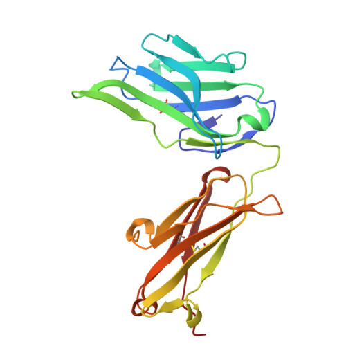 | |
Entity ID: 2 | |||||
|---|---|---|---|---|---|
| Molecule | Chains | Sequence Length | Organism | Details | Image |
| Anti-CD63 VL-CL iso | 218 | Homo sapiens | Mutation(s): 0 | 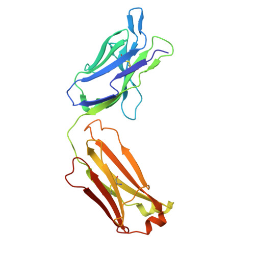 | |
| Length ( Å ) | Angle ( ˚ ) |
|---|---|
| a = 101.233 | α = 90 |
| b = 101.233 | β = 90 |
| c = 212.417 | γ = 90 |
| Software Name | Purpose |
|---|---|
| MOLREP | phasing |
| PHENIX | refinement |
| XDS | data reduction |
| XDS | data scaling |
| Coot | model building |
| Funding Organization | Location | Grant Number |
|---|---|---|
| Japan Society for the Promotion of Science (JSPS) | Japan | -- |


