


Crystal structure of cupin-domain containing protein (AzcA) from Streptomyces mobaraensis in complex with manganese and 6-amino-4-oxo-4,5-dihydro-1,3,5-triazine-2-carboxylic acid.
Nakashima, Y., Morita, H.To be published.
Experimental Data Snapshot
Starting Model: experimental
View more details
Entity ID: 1 | |||||
|---|---|---|---|---|---|
| Molecule | Chains | Sequence Length | Organism | Details | Image |
| Cupin type-2 domain-containing protein | 100 | Streptomyces mobaraensis | Mutation(s): 0 Gene Names: H7K43_04140 | 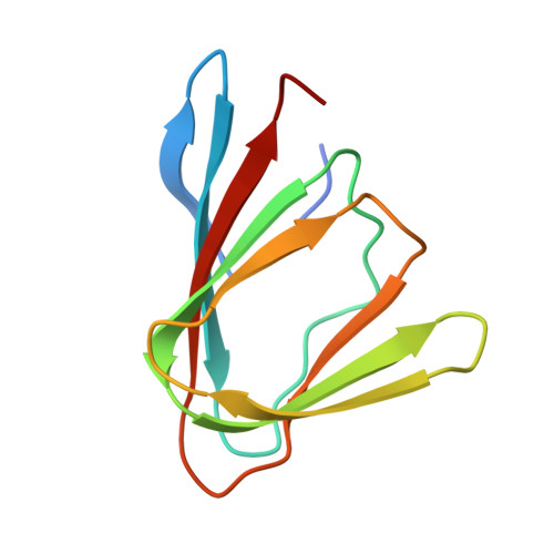 | |
| Ligands 4 Unique | |||||
|---|---|---|---|---|---|
| ID | Chains | Name / Formula / InChI Key | 2D Diagram | 3D Interactions | |
| A1L8W (Subject of Investigation/LOI) Download:Ideal Coordinates CCD File | F [auth A] | 2-azanyl-6-oxidanylidene-1~{H}-1,3,5-triazine-4-carboxylic acid C4 H4 N4 O3 OOQZQXLIGLORLI-UHFFFAOYSA-N |  | ||
| GOL (Subject of Investigation/LOI) Download:Ideal Coordinates CCD File | E [auth A], J [auth B] | GLYCEROL C3 H8 O3 PEDCQBHIVMGVHV-UHFFFAOYSA-N |  | ||
| IMD (Subject of Investigation/LOI) Download:Ideal Coordinates CCD File | H [auth B], I [auth B], L [auth C], N [auth D] | IMIDAZOLE C3 H5 N2 RAXXELZNTBOGNW-UHFFFAOYSA-O |  | ||
| MN (Subject of Investigation/LOI) Download:Ideal Coordinates CCD File | G [auth A], K [auth B], M [auth C], O [auth D] | MANGANESE (II) ION Mn WAEMQWOKJMHJLA-UHFFFAOYSA-N |  | ||
| Length ( Å ) | Angle ( ˚ ) |
|---|---|
| a = 58.523 | α = 90 |
| b = 72.024 | β = 90 |
| c = 97.401 | γ = 90 |
| Software Name | Purpose |
|---|---|
| PHENIX | refinement |
| XDS | data reduction |
| Aimless | data scaling |
| PHASER | phasing |
| Funding Organization | Location | Grant Number |
|---|---|---|
| Japan Society for the Promotion of Science (JSPS) | Japan | 22H04976 |
| Japan Society for the Promotion of Science (JSPS) | Japan | 18H03937 |
| Japan Society for the Promotion of Science (JSPS) | Japan | 22H05130 |


