 Download Mendeley
Standalone anti-phage HEPN nuclease PD-T7-3 is activated by ssDNA for tRNA cleavage
Puteikiene, R., Vassallo, C.N., Silanskas, A., Songailiene, I., Juozapaitis, J., Laub, M.T., Sasnauskas, G.To be published.
Experimental Data Snapshot
Starting Model: in silico
View more details
wwPDB Validation 3D Report Full Report

Entity ID: 1 | |||||
|---|---|---|---|---|---|
| Molecule | Chains | Sequence Length | Organism | Details | Image |
| Bacterial antiviral defense protein PD-T7-3 from Escherichia coli strain ECOR30 | 465 | Escherichia coli | Mutation(s): 0 | 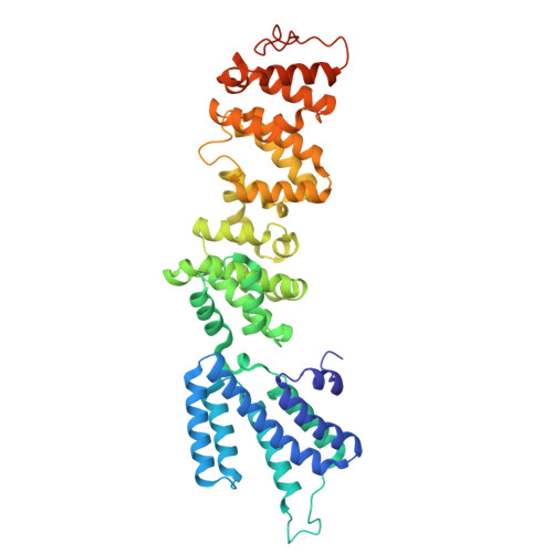 | |
UniProt | |||||
Entity Groups | |||||
| Sequence Clusters | 30% Identity50% Identity70% Identity90% Identity95% Identity100% Identity | ||||
| UniProt Group | Q858S9 | ||||
Sequence AnnotationsExpand | |||||
Reference Sequence | |||||
| Task | Software Package | Version |
|---|---|---|
| RECONSTRUCTION | cryoSPARC | 4.7.1 |
| MODEL REFINEMENT | PHENIX | 1.21.2-5419 |
| Funding Organization | Location | Grant Number |
|---|---|---|
| Research Council of Lithuania | Lithuania | S-MIP-22-13 |
