9T9P | pdb_00009t9p

Adenosine receptor A2a (A2AR)-beta-lactamase fusion bound to beta-lactamase inhibitory protein II (BLIPII) and ZM241385


Experimental Data Snapshot

  • Method: ELECTRON MICROSCOPY
  • Resolution: 3.00 Å
  • Aggregation State: PARTICLE 
  • Reconstruction Method: SINGLE PARTICLE 

Starting Models: experimental
View more details

wwPDB Validation 3D Report Full Report

Validation slider image for 9T9P

This is version 1.0 of the entry. See complete history

Literature

A novel fusion tool to enable G protein-coupled receptor structure determination.

Shah, N.R.Oosterlaken, M.Bisson, C.Bertinelli, M.Churchill-Angus, A.M.Hutchin, A.Kopec, J.Kotov, V.McFarlane, C.R.Griss Pascualli, E.Pavic, A.Zebisch, M.Fiez-Vandal, C.Fabini, E.Duclos, S.

(2026) Acta Crystallogr D Struct Biol 

  • DOI: https://doi.org/10.1107/S2059798326003785
  • Primary Citation Related Structures: 
    9T9P

  • PubMed Abstract: 

    Structure determination of G protein-coupled receptors (GPCRs) plays an important role in accelerating drug development against this medically important protein family. This study outlines the development of a new fusion tool to enable structure determination of GPCRs in inactive conformations by cryo-EM. Initially, a PDB mining approach was applied to select eight naturally occurring proteins with the intention of fusing them into the intracellular loop 3 (ICL3) of GPCRs to create a suitable fiducial marker for cryo-EM workflows. During the selection process, candidates with known high-affinity protein binders were prioritized to enable a further increase in the protein mass of the fiducial marker. Fusion constructs were generated with adenosine receptor A 2A (A 2A R) and were assessed for expression and aggregation levels. For the two best-performing new fusion constructs, ligand binding was characterized to ensure that the fusion tag did not significantly affect protein behaviour. A 2A R with a β-lactamase fusion in ICL3 and binding partner β-lactamase inhibitory protein II (BLIPII) was then selected to solve an antagonist-bound structure. The overall map was resolved to an average of 3.2 Å resolution with continuous helices connecting the β-lactamase to helices 5 and 6 of A 2A R. Focused refinement of the A 2A R region improved the local resolution and map detail in the orthosteric site, thereby allowing confident modelling of the antagonist ligand, ZM241385, which matches previously described X-ray crystallographic structures. This new fusion provides an alternative option for GPCR structure determination, with several potential benefits compared with existing tools, such as a more favourable position relative to the GPCR to reduce potential clashes.


  • Organizational Affiliation
    • Evotec (United Kingdom), Innovation Drive, Milton Park, Abingdon OX14 4RZ, United Kingdom.

Macromolecule Content 

  • Total Structure Weight: 124.37 kDa 
  • Atom Count: 6,115 
  • Modeled Residue Count: 803 
  • Deposited Residue Count: 1,133 
  • Unique protein chains: 2

Macromolecules

Find similar proteins by:|  3D Structure
Entity ID: 1
MoleculeChains  Sequence LengthOrganismDetailsImage
Beta-lactamase inhibitory protein II287Streptomyces exfoliatusMutation(s): 0 
Gene Names: bliB
UniProt
Find proteins for O87916 (Streptomyces exfoliatus)
Explore O87916 
Go to UniProtKB:  O87916
Entity Groups
Sequence Clusters30% Identity50% Identity70% Identity90% Identity95% Identity100% Identity
UniProt GroupO87916
Sequence Annotations
Expand
Reference Sequence
Find similar proteins by:|  3D Structure
Entity ID: 2
MoleculeChains  Sequence LengthOrganismDetailsImage
Adenosine receptor A2a,Small exopenicillinase,Green fluorescent protein846Homo sapiensBacillus licheniformisAequorea victoriaMutation(s): 0 
Gene Names: ADORA2AADORA2penPblaPGFP
EC: 3.5.2.6
UniProt & NIH Common Fund Data Resources
Find proteins for P29274 (Homo sapiens)
Explore P29274 
Go to UniProtKB:  P29274
PHAROS:  P29274
GTEx:  ENSG00000128271 
Find proteins for P00808 (Bacillus licheniformis)
Explore P00808 
Go to UniProtKB:  P00808
Find proteins for P42212 (Aequorea victoria)
Explore P42212 
Go to UniProtKB:  P42212
Entity Groups
Sequence Clusters30% Identity50% Identity70% Identity90% Identity95% Identity100% Identity
UniProt GroupsP42212P00808P29274
Sequence Annotations
Expand
Reference Sequence

Small Molecules

Ligands 1 Unique
IDChains Name / Formula / InChI Key2D Diagram3D Interactions
ZMA
(Subject of Investigation/LOI)

Query on ZMA


Download:Ideal Coordinates CCD File
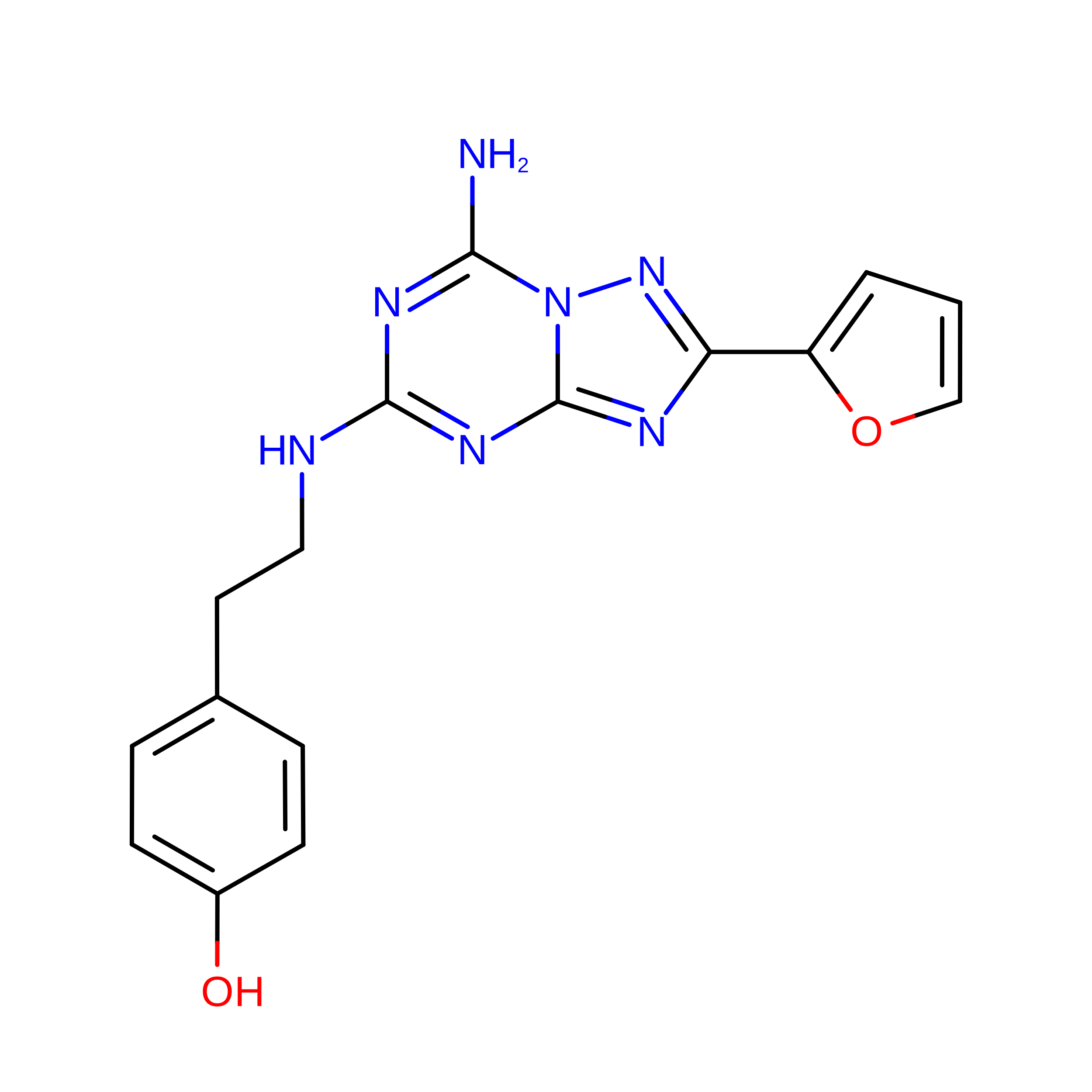
C [auth B]4-{2-[(7-amino-2-furan-2-yl[1,2,4]triazolo[1,5-a][1,3,5]triazin-5-yl)amino]ethyl}phenol
C16 H15 N7 O2
PWTBZOIUWZOPFT-UHFFFAOYSA-N

Experimental Data & Validation

Experimental Data

  • Method: ELECTRON MICROSCOPY
  • Resolution: 3.00 Å
  • Aggregation State: PARTICLE 
  • Reconstruction Method: SINGLE PARTICLE 
EM Software:
TaskSoftware PackageVersion
MODEL REFINEMENTPHENIX"2.0_5885"
RECONSTRUCTIONcryoSPARC4.7.1

Structure Validation

View Full Validation Report


Entry History 

& Funding Information

Deposition Data


Funding OrganizationLocationGrant Number
Not funded--

Revision History  (Full details and data files)

  • Version 1.0: 2026-05-27
    Type: Initial release