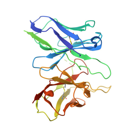
9T7P | pdb_00009t7p

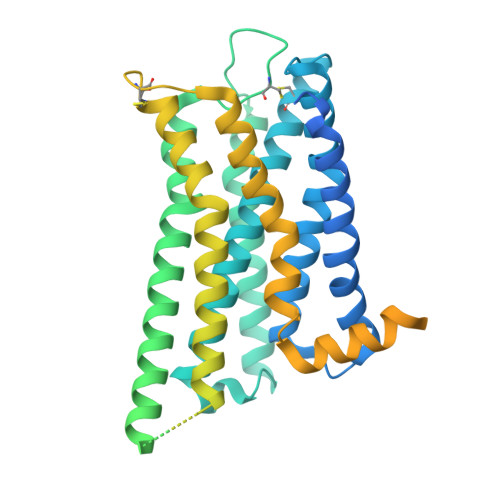
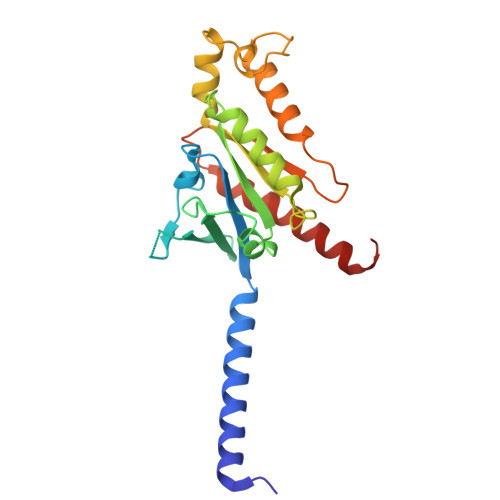
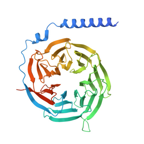
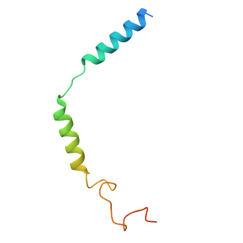
Cryo-EM structure of ESG-2-36-bound D3 dopamine receptor with mini-Go


Experimental Data Snapshot

  • Method: ELECTRON MICROSCOPY
  • Resolution: 2.85 Å
  • Aggregation State: PARTICLE 
  • Reconstruction Method: SINGLE PARTICLE 

wwPDB Validation 3D Report Full Report

Validation slider image for 9T7P

This is version 1.0 of the entry. See complete history

Literature

The structure of the dopamine D 3 receptor bound to cariprazine reveals principles for partial agonists with designed pharmacology.

Hadas Yardeni, E.Kiss, D.J.Sanchez, J.Shavit, K.Szepesi Kovacs, D.Egyed, A.Vogt, C.D.Gaitonde, S.A.Glenn, J.Canals, M.Bouvier, M.Newman, A.H.Lane, J.R.Keseru, G.M.Shalev-Benami, M.

(2026) Sci Adv 12: eaef7411-eaef7411

  • DOI: https://doi.org/10.1126/sciadv.aef7411
  • Primary Citation Related Structures: 
    9T7P

  • PubMed Abstract: 

    The third-generation antipsychotic cariprazine is a low-efficacy partial agonist of the dopamine D 3 receptor (D 3 R). Here, we report the cryo-electron microscopy structure of cariprazine bound to D 3 R, establishing a framework for understanding ligand recognition in this receptor. We further determine structures of D 3 R in complex with a series of cariprazine derivatives spanning inverse agonists to high-efficacy partial agonists. Integration of structural data with pharmacological profiling and molecular dynamics simulations reveals how subtle chemical modifications translate into distinct functional outcomes. Determinants distinguishing agonism from inverse agonism are well defined, whereas differences among partial agonists arise from small positional shifts of the ligand within the orthosteric binding site. In contrast, the extended binding site primarily modulates ligand stability, affinity, and receptor selectivity. These findings establish a mechanistic link between bitopic ligand architecture and receptor activation, providing a "ligand-centric" view of D 3 R signaling. Leveraging these principles, we designed and validated cariprazine derivatives with enhanced D 3 /D 2 selectivity and partial agonist activity. Together, this work provides a structural and pharmacological blueprint for the rational design of D 3 R-targeting ligands with tailored efficacy and therapeutic profiles.


  • Organizational Affiliation
    • Department of Chemical and Structural Biology, Weizmann Institute of Science, Rehovot, Israel.

Macromolecule Content 

  • Total Structure Weight: 167.71 kDa 
  • Atom Count: 8,605 
  • Modeled Residue Count: 1,120 
  • Deposited Residue Count: 1,531 
  • Unique protein chains: 5

Macromolecules

Find similar proteins by:|  3D Structure
Entity ID: 1
MoleculeChains  Sequence LengthOrganismDetailsImage
D(3) dopamine receptor,LgbitA [auth R]594Homo sapiensMutation(s): 0 
Gene Names: DRD3
UniProt & NIH Common Fund Data Resources
Find proteins for P35462 (Homo sapiens)
Explore P35462 
Go to UniProtKB:  P35462
PHAROS:  P35462
GTEx:  ENSG00000151577 
Entity Groups
Sequence Clusters30% Identity50% Identity70% Identity90% Identity95% Identity100% Identity
UniProt GroupP35462
Sequence Annotations
Expand
Reference Sequence
Find similar proteins by:|  3D Structure
Entity ID: 2
MoleculeChains  Sequence LengthOrganismDetailsImage
Engineered miniGo,Guanine nucleotide-binding protein G(o) subunit alpha,Guanine nucleotide-binding protein G(o) subunit alphaB [auth A]226Homo sapiensMutation(s): 0 
EC: 3.6.5
UniProt & NIH Common Fund Data Resources
Find proteins for P09471 (Homo sapiens)
Explore P09471 
Go to UniProtKB:  P09471
PHAROS:  P09471
GTEx:  ENSG00000087258 
Entity Groups
Sequence Clusters30% Identity50% Identity70% Identity90% Identity95% Identity100% Identity
UniProt GroupP09471
Sequence Annotations
Expand
Reference Sequence
Find similar proteins by:|  3D Structure
Entity ID: 3
MoleculeChains  Sequence LengthOrganismDetailsImage
Guanine nucleotide-binding protein G(I)/G(S)/G(T) subunit beta-1C [auth B]384Homo sapiensMutation(s): 0 
Gene Names: GNB1
UniProt & NIH Common Fund Data Resources
Find proteins for P62873 (Homo sapiens)
Explore P62873 
Go to UniProtKB:  P62873
PHAROS:  P62873
GTEx:  ENSG00000078369 
Entity Groups
Sequence Clusters30% Identity50% Identity70% Identity90% Identity95% Identity100% Identity
UniProt GroupP62873
Sequence Annotations
Expand
Reference Sequence
Find similar proteins by:|  3D Structure
Entity ID: 4
MoleculeChains  Sequence LengthOrganismDetailsImage
Guanine nucleotide-binding protein G(I)/G(S)/G(O) subunit gamma-2D [auth C]71Homo sapiensMutation(s): 0 
Gene Names: GNG2
UniProt & NIH Common Fund Data Resources
Find proteins for P59768 (Homo sapiens)
Explore P59768 
Go to UniProtKB:  P59768
PHAROS:  P59768
GTEx:  ENSG00000186469 
Entity Groups
Sequence Clusters30% Identity50% Identity70% Identity90% Identity95% Identity100% Identity
UniProt GroupP59768
Sequence Annotations
Expand
Reference Sequence
Find similar proteins by:|  3D Structure
Entity ID: 5
MoleculeChains  Sequence LengthOrganismDetailsImage
scFv16E [auth S]256Mus musculusMutation(s): 0 
Entity Groups
Sequence Clusters30% Identity50% Identity70% Identity90% Identity95% Identity100% Identity
Sequence Annotations
Expand
Reference Sequence

Small Molecules

Ligands 1 Unique
IDChains Name / Formula / InChI Key2D Diagram3D Interactions
A1JUA(
Subject of Investigation/LOI)

Query on A1JUA


Download:Ideal Coordinates CCD File
F [auth R]3-[4-[2-[4-(2-fluoranyl-3-methoxy-phenyl)piperazin-1-yl]ethyl]cyclohexyl]-1,1-dimethyl-urea
C22 H35 F N4 O2
MUUSXXFKLPMJKL-IYARVYRRSA-N

Experimental Data & Validation

Experimental Data

  • Method: ELECTRON MICROSCOPY
  • Resolution: 2.85 Å
  • Aggregation State: PARTICLE 
  • Reconstruction Method: SINGLE PARTICLE 
EM Software:
TaskSoftware PackageVersion
RECONSTRUCTIONcryoSPARC

Structure Validation

View Full Validation Report


Entry History 

& Funding Information

Deposition Data


Funding OrganizationLocationGrant Number
European Research Council (ERC)European Union949364

Revision History  (Full details and data files)

  • Version 1.0: 2026-09-09
    Type: Initial release