Engineering SIRP alpha conformational plasticity to reveal a cryptic pocket suitable for structure-based drug design.
Storder, M., Barelier, S., Cordier, F., Yacoub, T., Ilari, L., Barral, K., Mahmoodi, S., Saez-Ayala, M., Combes, S., Betzi, S., Derviaux, C., Ulliana, A., Torres, F., Rubin, J., Roche, P., Morelli, X., Garcin, E.D., Miller, T.W.(2025) bioRxiv
- PubMed: 41497624 Search on PubMedSearch on PubMed Central
- DOI: https://doi.org/10.64898/2025.12.10.693509
- Primary Citation Related Structures:
9SIA, 9SIC, 9SID, 9T7F, 9TF5 - PubMed Abstract:
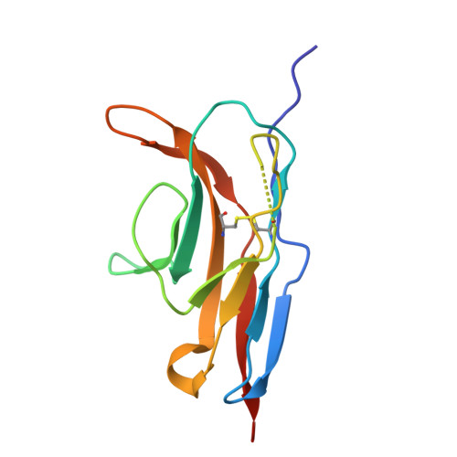
The protein-protein interaction between Signal Regulatory Protein alpha (SIRPα) and CD47 is a critical immune checkpoint that enables tumor immune escape, making it a key target for cancer immunotherapy. While antibody-based therapies exist, the development of small-molecule inhibitors has been hindered by the flat, featureless binding interface. Here, we report the discovery of a novel, druggable cryptic pocket within the SIRPα D1 domain (the WYF pocket), revealed through a structure-based fragment screening campaign using x-ray crystallography. This pocket, defined by residues Trp38, Tyr50, and Phe74, is only accessible in a conformation that is incompatible with CD47 binding, making it a candidate for structure-based drug design and immune checkpoint inhibitor development. Through a combination of NMR spectroscopy, molecular dynamics simulations, and biophysical assays, we demonstrate that access to this cryptic site is dynamically controlled by a single "gatekeeper" residue, Gln52. The rotameric state of Gln52 dictates a conformational equilibrium between a "closed," state and a ligand-accessible "open" state. We validated this mechanism by engineering SIRPα mutants to bias this equilibrium. A Q52F mutation locked the pocket in a closed state, abolishing both CD47 and fragment binding, while Q52A and Q52R mutations biased the protein toward an open state. These "open-biased" mutants not only exhibited decreased affinity for CD47 but also significantly improved binding to small-molecule fragments that inhibit the SIRPα-CD47 interaction. This work reveals the intrinsic conformational plasticity of SIRPα and establishes a validated structure-based roadmap for a new class of allosteric inhibitors. This 'flexibility-for-inhibition' strategy functions by trapping a non-binding conformation and represents a broadly applicable framework for targeting this and other challenging immune checkpoints.
- CRCM, CNRS, Inserm, Institut Paoli-Calmettes, Aix-Marseille Univ, Marseille, France.
Organizational Affiliation: