Download Mendeley
Structures of the volume-regulated anion channel LRRC8A/D in activating and inhibiting conditions.
Lehmann, E.F., Stierli, F., Rutz, S., Deneka, D., Dutzler, R.(2026) Nat Commun 17
- PubMed: 42754598 Search on PubMedSearch on PubMed Central
- DOI: https://doi.org/10.1038/s41467-026-77508-x
- Primary Citation Related Structures:
9T5M, 9T5N, 9T5O, 9T5P, 9T5Q, 9T5R - PubMed Abstract:
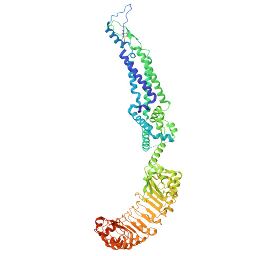
Volume-regulated anion channels, which are activated in response to hypotonicity, are heteromers composed of LRRC8 family members. They consist of the obligatory subunit LRRC8A and at least another paralog that determines their substrate preference. Here we investigate heteromeric assemblies composed of LRRC8A and D subunits, the latter of which confer permeability to amino acids, osmolytes and anti-cancer drugs. Using patch-clamp electrophysiology, we show that LRRC8A/D channels are reversibly activated by cell swelling and that their response is modulated by sybodies targeting the A subunits, which either potentiate or repress channel activity. Structures of the LRRC8A/D channel in complex with the inhibitory sybody Sb1 define the channel stoichiometry of four A and two D subunits, with Sb1 stabilizing the A subunits in a similar closed channel conformation as found in homomeric LRRC8A assemblies. In LRRC8A/D heteromers, the two D subunits weaken the arrangement of A subunits and thus enhance the activation properties of the channel. This channel conformation is strongly perturbed upon binding of the potentiating sybody Sb4, which disrupts the packing of the cytoplasmic domains linking their mobility to pore opening.
- Department of Biochemistry University of Zurich, Winterthurerstrasse 190, Zurich, Switzerland.
Organizational Affiliation: