Download Mendeley
Spatiotemporal expression of endospore appendages and cryo-EM insights into Ena1C-mediated S-ENA anchoring in Bacillus paranthracis.
Zegeye, E.D., Sleutel, M., Jonsmoen, U.L., Chen, J., Morawska, L.P., Mekonnen, Y.B., Kuipers, O.P., Remaut, H., Aspholm, M.(2026) Sci Rep 16: 7122-7122
- PubMed: 41634327 Search on PubMedSearch on PubMed Central
- DOI: https://doi.org/10.1038/s41598-026-38321-0
- Primary Citation Related Structures:
9T3P - PubMed Abstract:
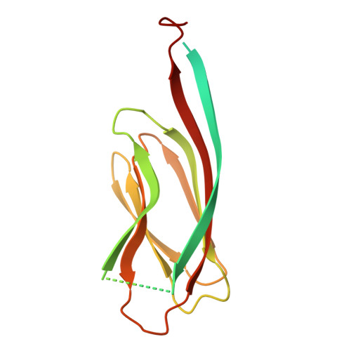
The endospores of many Bacillus cereus group species are decorated with highly resilient fibrous structures known as endospore appendages (ENAs), whose precise biological functions remain poorly understood. Structural and genetic studies have identified ena1A, ena1B, and ena1C as essential for forming the longer, thicker, and most abundant staggered (S)-ENA fibers in Bacillus paranthracis, whereas ena3A encodes the major subunit of the shorter, thinner, ladder-like (L)-ENAs. Here, we investigated the spatiotemporal expression dynamics of S- and L-ENA proteins and the specific role of Ena1C in S-ENA biogenesis. Using time-lapse fluorescence microscopy, we observed strict temporal regulation of ena gene expression, with no detectable ENA subunit production before spores became phase-bright. ENAs expression peaked during late sporulation phase, with fluorescence localized around the developing spore until its release; notably, S-ENA subunit expression began approximately one hour earlier than that of L-ENA subunits. Combining cryo-EM, negative-stain transmission electron microscopy, and genetic analyses, we show that Ena1C forms a nonameric ring-like structure required for tethering S-ENA to the spore surface. These findings provide new insights into the regulation of ENAs’ expression during fiber biogenesis and highlight their temporal coordination with spore coat and exosporium development.
- Department of Paraclinical Sciences, Faculty of Veterinary Medicine , Norwegian University of Life Sciences (NMBU) , Ås, 1433, Norway. ephrem.debebe.zegeye@nmbu.no.
Organizational Affiliation: