9T09 | pdb_00009t09

X-ray structure of the catalytic domain of CDC25B C473S at 1.34 angstrom resolution


Experimental Data Snapshot

  • Method: X-RAY DIFFRACTION
  • Resolution: 1.34 Å
  • R-Value Free: 
    0.175 (Depositor), 0.175 (DCC) 
  • R-Value Work: 
    0.157 (Depositor), 0.157 (DCC) 
  • R-Value Observed: 
    0.158 (Depositor) 

Starting Model: experimental
View more details

wwPDB Validation 3D Report Full Report

Validation slider image for 9T09

This is version 1.1 of the entry. See complete history

Literature

The Binding of 3-O-Methylfluorescein Phosphate to the Catalytic Domain of the Human CDC25B Phosphatase: A Structural Investigation.

Troisi, R.Rullo, R.Napolitano, V.Popowicz, G.M.De Vendittis, E.Sica, F.

(2026) Chembiochem 27: e202600010-e202600010

  • DOI: https://doi.org/10.1002/cbic.202600010
  • Primary Citation Related Structures: 
    9T09, 9T0A

  • PubMed Abstract: 

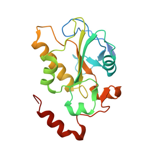
    The molecular mechanisms by which the human CDC25B activates the CDK1/cyclin B complex in the cell cycle, as well as how it can be inhibited by synthetic inhibitors at the atomic level, are still under investigation. Valuable insights have been gained from the molecular structure here-described, which captures for the first time the interaction between the C-terminal domain of the inactive mutant CDC25B C473S (CDC25B-S) and the commonly used synthetic substrate 3-O-methylfluorescein phosphate (3-OMFP). Crystallographic studies reveal that 3-OMFP engages multiple residues within the active site and the adjacent "swimming pool" of CDC25B-S, establishing specific interactions and prompting local adjustments in this region. These structural features explain the increased resistance to thermal denaturation of CDC25B-S observed through circular dichroism measurements upon substrate binding. The structural changes induced by 3-OMFP lead to a conformation comparable to that of CDC25A bound to its substrate, the CDK2/cyclin A complex. These findings qualify 3-OMFP as a promising starting model for the rational design of selective competitive inhibitors of CDC25B having reduced off-target effects.


  • Organizational Affiliation
    • Department of Chemical Sciences, University of Naples Federico II, Complesso Universitario di Monte Sant'Angelo, Naples, Italy.

Macromolecule Content 

  • Total Structure Weight: 23.76 kDa 
  • Atom Count: 1,939 
  • Modeled Residue Count: 180 
  • Deposited Residue Count: 197 
  • Unique protein chains: 1

Macromolecules

Find similar proteins by:|  3D Structure
Entity ID: 1
MoleculeChains  Sequence LengthOrganismDetailsImage
M-phase inducer phosphatase 2197Homo sapiensMutation(s): 1 
Gene Names: CDC25BCDC25HU2
EC: 3.1.3.48
UniProt & NIH Common Fund Data Resources
Find proteins for P30305 (Homo sapiens)
Explore P30305 
Go to UniProtKB:  P30305
PHAROS:  P30305
GTEx:  ENSG00000101224 
Entity Groups
Sequence Clusters30% Identity50% Identity70% Identity90% Identity95% Identity100% Identity
UniProt GroupP30305
Sequence Annotations
Expand
Reference Sequence

Small Molecules

Ligands 4 Unique
IDChains Name / Formula / InChI Key2D Diagram3D Interactions
SO4

Query on SO4


Download:Ideal Coordinates CCD File
B [auth A],
C [auth A],
D [auth A],
E [auth A]
SULFATE ION
O4 S
QAOWNCQODCNURD-UHFFFAOYSA-L
GOL

Query on GOL


Download:Ideal Coordinates CCD File
H [auth A],
I [auth A],
J [auth A]
GLYCEROL
C3 H8 O3
PEDCQBHIVMGVHV-UHFFFAOYSA-N
ZN

Query on ZN


Download:Ideal Coordinates CCD File
G [auth A]ZINC ION
Zn
PTFCDOFLOPIGGS-UHFFFAOYSA-N
CL

Query on CL


Download:Ideal Coordinates CCD File
F [auth A]CHLORIDE ION
Cl
VEXZGXHMUGYJMC-UHFFFAOYSA-M

Experimental Data & Validation

Experimental Data

  • Method: X-RAY DIFFRACTION
  • Resolution: 1.34 Å
  • R-Value Free:  0.175 (Depositor), 0.175 (DCC) 
  • R-Value Work:  0.157 (Depositor), 0.157 (DCC) 
  • R-Value Observed: 0.158 (Depositor) 
Space Group: P 21 21 21
Unit Cell:
Length ( Å )Angle ( ˚ )
a = 52.13α = 90
b = 71.2β = 90
c = 72.9γ = 90
Software Package:
Software NamePurpose
XDSdata reduction
XSCALEdata scaling
autoPROCdata processing
PHASERphasing
REFMACrefinement
PDB_EXTRACTdata extraction

Structure Validation

View Full Validation Report


Entry History 

& Funding Information

Deposition Data


Funding OrganizationLocationGrant Number
Not funded--

Revision History  (Full details and data files)

  • Version 1.0: 2026-03-11
    Type: Initial release
  • Version 1.1: 2026-04-08
    Changes: Database references