9SSM | pdb_00009ssm

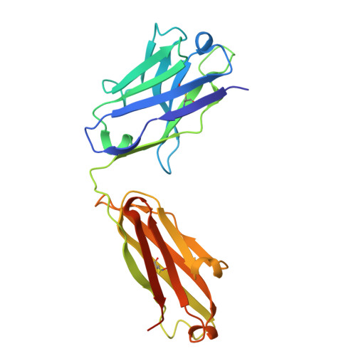
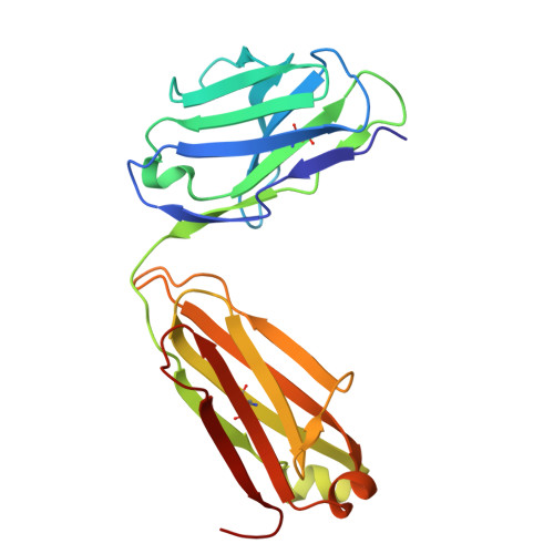
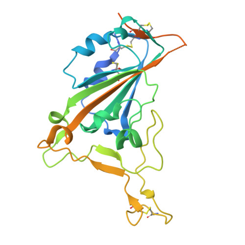
Crystal structure of 084-7D Fab bound to SARS-CoV-2 Beta RBD


Experimental Data Snapshot

  • Method: X-RAY DIFFRACTION
  • Resolution: 2.70 Å
  • R-Value Free: 
    0.269 (Depositor), 0.267 (DCC) 
  • R-Value Work: 
    0.223 (Depositor), 0.222 (DCC) 
  • R-Value Observed: 
    0.225 (Depositor) 

Starting Model: in silico
View more details

wwPDB Validation 3D Report Full Report

Validation slider image for 9SSM

Ligand Structure Quality Assessment 


This is version 1.1 of the entry. See complete history

Literature

Defining the mechanism of cross-reactivity for a SARS-CoV-2 Beta-elicited antibody toward omicron sub-lineages.

Ayres, F.Lambson, B.Mkhize, N.N.Makhado, Z.Mhlanga, D.Serage, R.Moore, P.L.Wibmer, C.K.Moyo-Gwete, T.

(2026) Structure 34: 552

  • DOI: https://doi.org/10.1016/j.str.2026.01.006
  • Primary Citation Related Structures: 
    9SSM

  • PubMed Abstract: 

    Despite the continual emergence of SARS-CoV-2 variants and increasing diversity within the receptor binding domain (RBD), some antibody responses that are directed to conserved regions can display cross-reactivity against variants. We previously isolated an RBD-directed monoclonal antibody (084-7D) from a Beta-infected donor that neutralized Beta and emerging Omicron variants. Here, we solved a high-resolution crystal structure of the 084-7D Fab in complex with the Beta RBD. These data revealed an epitope overlapping both the ACE2 binding site and those of other class 1 antibodies. Furthermore, the epitope includes highly conserved residues, Q409, D420, and Y489, that are present in recent Omicron variants. The N417 residue that emerged with Beta and has since persisted is tolerated within the epitope of 084-7D, explaining the preferential neutralization of contemporaneous N417-containing variants. These structural data defined the mechanism for cross-reactivity of a Beta-elicited neutralizing antibody, potentially informing the design of future broadly reactive SARS-CoV-2 therapeutics.


  • Organizational Affiliation
    • SA MRC Antibody Immunity Research Unit, School of Pathology, Faculty of Health Sciences, University of the Witwatersrand, Johannesburg, South Africa; Wits Health Consortium, University of the Witwatersrand, Johannesburg, South Africa; Venom & Vaccine Immunotherapeutic & Immunogen Protein Engineering Research (VïPER), Department of Integrative Biomedical Sciences, Faculty of Health Sciences, University of Cape Town, Cape Town, South Africa; National Institute for Communicable Diseases of the National Health Laboratory Service, Johannesburg, South Africa.

Macromolecule Content 

  • Total Structure Weight: 76.16 kDa 
  • Atom Count: 4,717 
  • Modeled Residue Count: 626 
  • Deposited Residue Count: 693 
  • Unique protein chains: 3

Macromolecules

Find similar proteins by:|  3D Structure
Entity ID: 1
MoleculeChains  Sequence LengthOrganismDetailsImage
084-7D Fab Heavy ChainA [auth H]230Homo sapiensMutation(s): 0 
Gene Names: IgHV
Find similar proteins by:|  3D Structure
Entity ID: 2
MoleculeChains  Sequence LengthOrganismDetailsImage
084-7D Fab Light ChainB [auth K]214Homo sapiensMutation(s): 0 
Gene Names: IgKV
Find similar proteins by:|  3D Structure
Entity ID: 3
MoleculeChains  Sequence LengthOrganismDetailsImage
Spike glycoproteinC [auth R]249Severe acute respiratory syndrome coronavirus 2Mutation(s): 0 
UniProt
Find proteins for A0A8A5XRG7 (Severe acute respiratory syndrome coronavirus 2)
Explore A0A8A5XRG7 
Go to UniProtKB:  A0A8A5XRG7
Entity Groups
Sequence Clusters30% Identity50% Identity70% Identity90% Identity95% Identity100% Identity
UniProt GroupA0A8A5XRG7
Glycosylation
Glycosylation Sites: 1
Sequence Annotations
Expand
Reference Sequence

Small Molecules

Ligands 1 Unique
IDChains Name / Formula / InChI Key2D Diagram3D Interactions
NAG

Query on NAG


Download:Ideal Coordinates CCD File
D [auth R]2-acetamido-2-deoxy-beta-D-glucopyranose
C8 H15 N O6
OVRNDRQMDRJTHS-FMDGEEDCSA-N

Experimental Data & Validation

Experimental Data

  • Method: X-RAY DIFFRACTION
  • Resolution: 2.70 Å
  • R-Value Free:  0.269 (Depositor), 0.267 (DCC) 
  • R-Value Work:  0.223 (Depositor), 0.222 (DCC) 
  • R-Value Observed: 0.225 (Depositor) 
Space Group: P 21 21 21
Unit Cell:
Length ( Å )Angle ( ˚ )
a = 81.45α = 90
b = 84.368β = 90
c = 126.239γ = 90
Software Package:
Software NamePurpose
PHENIXmodel building
PHENIXrefinement
xia2data scaling
xia2data reduction
PHASERphasing
PDB_EXTRACTdata extraction

Structure Validation

View Full Validation Report


Ligand Structure Quality Assessment 


Entry History 

& Funding Information

Deposition Data


Funding OrganizationLocationGrant Number
Wellcome TrustUnited Kingdom226137/Z/22/Z
European Union (EU)European Union101046041
South African Medical Research Council self-initiated research grant (SAMRC SIR Grant)South Africa--

Revision History  (Full details and data files)

  • Version 1.0: 2026-02-18
    Type: Initial release
  • Version 1.1: 2026-04-15
    Changes: Database references