9SOP | pdb_00009sop

Tissue inhibitor of metalloproteinase-1 (TIMP-1)

  • Classification: HYDROLASE
  • Organism(s): Homo sapiens
  • Expression System: Homo sapiens
  • Mutation(s): No 

  • Deposited: 2025-09-15 Released: 2025-09-15 
  • Deposition Author(s): Shemy, A., Voet, A.
  • Funding Organization(s): Research Foundation - Flanders (FWO)

Experimental Data Snapshot

  • Method: X-RAY DIFFRACTION
  • Resolution: 1.95 Å
  • R-Value Free: 
    0.239 (Depositor), 0.239 (DCC) 
  • R-Value Work: 
    0.194 (Depositor), 0.194 (DCC) 

Starting Model: experimental
View more details

wwPDB Validation 3D Report Full Report

Validation slider image for 9SOP

Ligand Structure Quality Assessment 


This is version 2.0 of the entry. See complete history

Literature

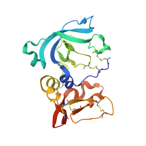
The human TIMP-1 unbound structure provides a platform for fragment screening.

Shemy, A.Van Broeckhoven, J.Hellings, N.Voet, A.

(2026) Acta Crystallogr D Struct Biol 82: 330-335

  • DOI: https://doi.org/10.1107/S2059798326001749
  • Primary Citation Related Structures: 
    9SOP, 9SOQ, 9SOS

  • PubMed Abstract: 

    Tissue inhibitor of metalloproteinases-1 (TIMP-1) is a critical regulator of extracellular matrix remodelling and an important mediator of remyelination in demyelinating disorders such as multiple sclerosis. In addition, TIMP-1 has emerged as a promising therapeutic target in cancer due to its interaction with CD63, which promotes tumorigenic signalling and carcinogenesis. Although several structures of TIMP-1 bound to matrix metalloproteinases have been reported, no unbound structure with all druggable sites available has previously been reported. Here, we present the first unbound crystal structure of human TIMP-1, resolved at 1.95 Å resolution. Comparison with the MMP-bound complex reveals localized conformational changes and altered intramolecular hydrogen bonding in the unbound structure, indicating increased structural plasticity in the absence of the protease. Crystals were obtained in multiple conditions, but only two diffracted to high resolution. Although optimization and seeding did not significantly improve the morphology, the additive screen enhanced both the morphology and reproducibility and provided intrinsic cryoprotection. The resulting crystal form proved compatible with soaking-based screening campaigns, providing a robust structural basis for the discovery of TIMP-1 ligands with clinical potential.


  • Organizational Affiliation
    • Biomolecular Modelling and Design Lab, Department of Chemistry, University of Leuven, Celestijnenlaan 200G, 3001 Heverlee, Belgium.

Macromolecule Content 

  • Total Structure Weight: 42.03 kDa 
  • Atom Count: 2,995 
  • Modeled Residue Count: 356 
  • Deposited Residue Count: 360 
  • Unique protein chains: 1

Macromolecules

Find similar proteins by:|  3D Structure
Entity ID: 1
MoleculeChains  Sequence LengthOrganismDetailsImage
Metalloproteinase inhibitor 1
A, B
180Homo sapiensMutation(s): 0 
Gene Names: TIMP1CLGITIMP
UniProt & NIH Common Fund Data Resources
Find proteins for P01033 (Homo sapiens)
Explore P01033 
Go to UniProtKB:  P01033
PHAROS:  P01033
GTEx:  ENSG00000102265 
Entity Groups
Sequence Clusters30% Identity50% Identity70% Identity90% Identity95% Identity100% Identity
UniProt GroupP01033
Sequence Annotations
Expand
Reference Sequence

Small Molecules

Ligands 5 Unique
IDChains Name / Formula / InChI Key2D Diagram3D Interactions
P3G

Query on P3G


Download:Ideal Coordinates CCD File
I [auth A]3,6,9,12,15-PENTAOXAHEPTADECANE
C12 H26 O5
HYDWALOBQJFOMS-UHFFFAOYSA-N
FLC

Query on FLC


Download:Ideal Coordinates CCD File
H [auth A]CITRATE ANION
C6 H5 O7
KRKNYBCHXYNGOX-UHFFFAOYSA-K
PGE

Query on PGE


Download:Ideal Coordinates CCD File
O [auth B]TRIETHYLENE GLYCOL
C6 H14 O4
ZIBGPFATKBEMQZ-UHFFFAOYSA-N
GOL

Query on GOL


Download:Ideal Coordinates CCD File
C [auth A],
F [auth A],
P [auth B]
GLYCEROL
C3 H8 O3
PEDCQBHIVMGVHV-UHFFFAOYSA-N
EDO

Query on EDO


Download:Ideal Coordinates CCD File
D [auth A]
E [auth A]
G [auth A]
J [auth B]
K [auth B]
D [auth A],
E [auth A],
G [auth A],
J [auth B],
K [auth B],
L [auth B],
M [auth B],
N [auth B]
1,2-ETHANEDIOL
C2 H6 O2
LYCAIKOWRPUZTN-UHFFFAOYSA-N

Experimental Data & Validation

Experimental Data

  • Method: X-RAY DIFFRACTION
  • Resolution: 1.95 Å
  • R-Value Free:  0.239 (Depositor), 0.239 (DCC) 
  • R-Value Work:  0.194 (Depositor), 0.194 (DCC) 
Space Group: P 1 21 1
Unit Cell:
Length ( Å )Angle ( ˚ )
a = 49.229α = 90
b = 44.241β = 99.06
c = 90.53γ = 90
Software Package:
Software NamePurpose
REFMACrefinement
REFMACrefinement
Aimlessdata scaling
DIALSdata reduction
gemmidata extraction
Aimlessdata scaling
DIALSdata reduction
gemmidata extraction
PHASERphasing

Structure Validation

View Full Validation Report


Ligand Structure Quality Assessment 


Entry History 

& Funding Information

Deposition Data


Funding OrganizationLocationGrant Number
Research Foundation - Flanders (FWO)Belgium1S11123N
Research Foundation - Flanders (FWO)Belgium1S11125N

Revision History  (Full details and data files)

  • Version 1.0: 2025-09-15
    Type: Initial release
  • Version 2.0: 2026-04-29
    Type: Coordinate replacement
    Reason: Ligand identity
    Changes: Advisory, Atomic model, Data collection, Database references, Derived calculations, Non-polymer description, Polymer sequence, Refinement description, Source and taxonomy, Structure summary