9SMY | pdb_00009smy

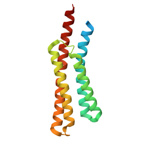
Structure of the ligand binding domain of the Pseudomonas putida chemoreceptor PcpI


Experimental Data Snapshot

  • Method: X-RAY DIFFRACTION
  • Resolution: 2.20 Å
  • R-Value Free: 
    0.234 (Depositor), 0.233 (DCC) 
  • R-Value Work: 
    0.186 (Depositor), 0.186 (DCC) 
  • R-Value Observed: 
    0.188 (Depositor) 

Starting Model: in silico
View more details

wwPDB Validation 3D Report Full Report

Validation slider image for 9SMY

This is version 1.0 of the entry. See complete history

Literature

Evolution of monomodular all-helical receptor ligand-binding domains from bimodular ancestors.

Gavira, J.A.Rico-Jimenez, M.Ortega, A.Roca, A.Krell, T.Zhulin, I.B.Matilla, M.A.

(2026) Int J Biol Macromol : 154135-154135

  • DOI: https://doi.org/10.1016/j.ijbiomac.2026.154135
  • Primary Citation Related Structures: 
    9SMY, 9SNN, 9SOU, 9SOY

  • PubMed Abstract: 

    Bacterial chemotaxis is essential for environmental adaptation and host interaction. To this end, bacteria have evolved exceptionally broad chemosensory capacities, with few apparent constraints on ligand structure or size. These capacities are determined by the extraordinary diversity of chemoreceptor ligand-binding domains (LBDs), which recognize chemoeffectors and evolve rapidly to acquire new functions. Many LBDs have complex architectures, often comprising multiple ligand-binding modules. Among chemoreceptor LBDs, members of the all-helical class are widespread and can contain one, two, or three stacked four-helix bundle (4HB) modules. Here, using phylogenomic, structural, and biochemical approaches, we identify a novel monomodular all-helical LBD family, termed 4HB_HD (4HB_HBM-derived), most likely originated from the bimodular all-helical HBM LBD by the loss of its membrane-distal module. A representative family member, PcpI of Pseudomonas putida, binds the plant hormones salicylate and indole-3-acetic acid and mediates chemotaxis toward these compounds. Comparison with the inferred bimodular ancestor, aPcpI, revealed that binds the phytohormones recognized by PcpI via both the membrane-distal and membrane-proximal modules, and additionally recognizes citrate through the membrane-distal module. Despite their distinct chemical structures, these ligands bind to the same site within the membrane-distal module, highlighting structural flexibility as a mechanism for expanding receptor specificity. Structural analyses further show that PcpI-LBD closely superimposes with the membrane-proximal module of the aPcpI-LBD and provide a structural rationale for its inability to bind citrate. Together, our results show that modular reduction does not necessarily compromise function and illustrate how rearrangement of ligand-binding modules can drive the microbial evolution of inter-kingdom signal detection.


  • Organizational Affiliation
    • Laboratory of Crystallographic Studies, Instituto Andaluz de Ciencias de la Tierra-Consejo Superior de Investigaciones Científicas, Armilla, 18100, Spain.

Macromolecule Content 

  • Total Structure Weight: 36.15 kDa 
  • Atom Count: 2,409 
  • Modeled Residue Count: 264 
  • Deposited Residue Count: 316 
  • Unique protein chains: 1

Macromolecules

Find similar proteins by:|  3D Structure
Entity ID: 1
MoleculeChains  Sequence LengthOrganismDetailsImage
HAMP domain-containing protein
A, B
158Pseudomonas putidaMutation(s): 0 
Gene Names: E6B08_28110
UniProt
Find proteins for A0A4D6XHR0 (Pseudomonas putida)
Explore A0A4D6XHR0 
Go to UniProtKB:  A0A4D6XHR0
Entity Groups
Sequence Clusters30% Identity50% Identity70% Identity90% Identity95% Identity100% Identity
UniProt GroupA0A4D6XHR0
Sequence Annotations
Expand
Reference Sequence

Small Molecules

Ligands 4 Unique
IDChains Name / Formula / InChI Key2D Diagram3D Interactions
PG4

Query on PG4


Download:Ideal Coordinates CCD File
I [auth A]TETRAETHYLENE GLYCOL
C8 H18 O5
UWHCKJMYHZGTIT-UHFFFAOYSA-N
PGE

Query on PGE


Download:Ideal Coordinates CCD File
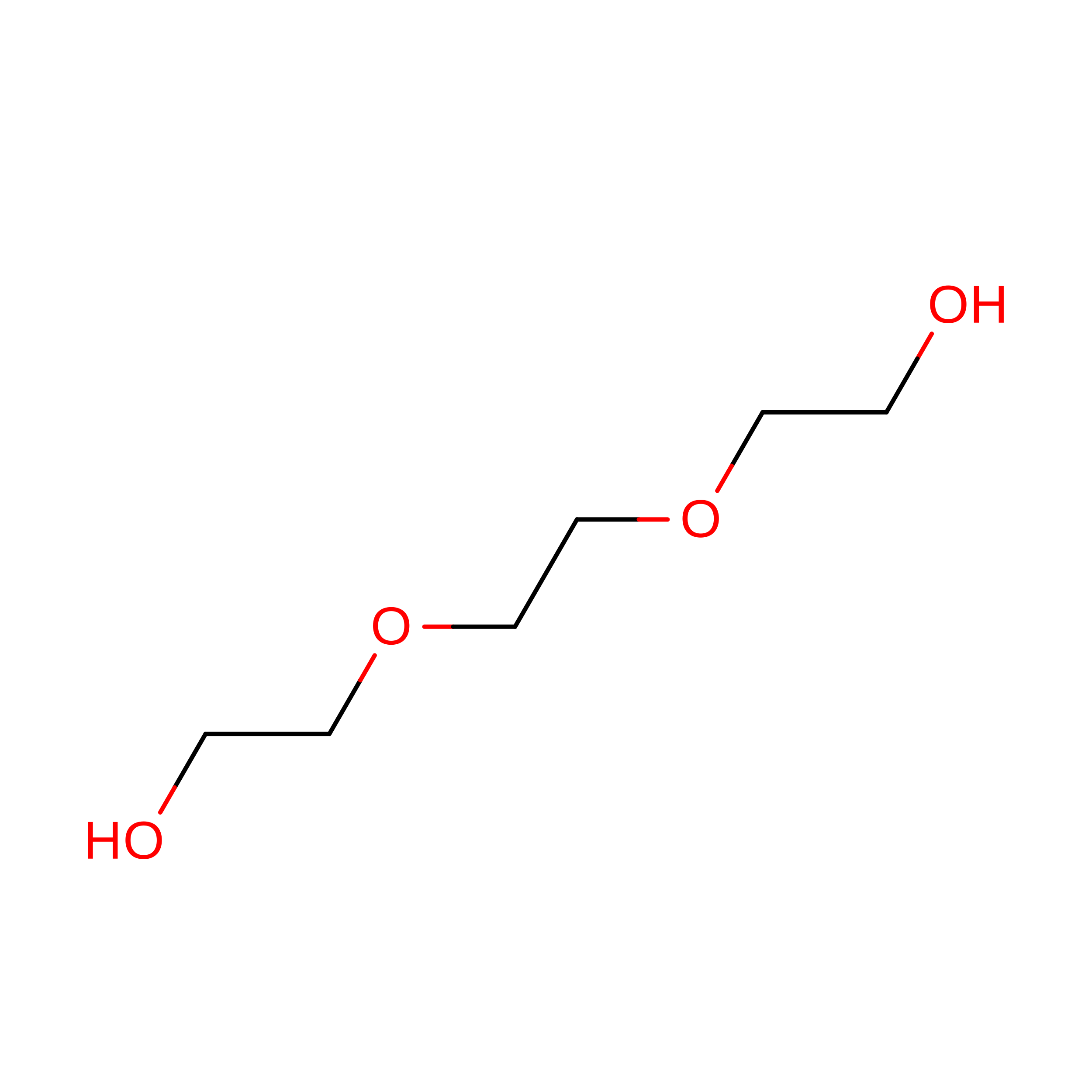
N [auth B]TRIETHYLENE GLYCOL
C6 H14 O4
ZIBGPFATKBEMQZ-UHFFFAOYSA-N
GOL

Query on GOL


Download:Ideal Coordinates CCD File
E [auth A],
G [auth A],
H [auth A]
GLYCEROL
C3 H8 O3
PEDCQBHIVMGVHV-UHFFFAOYSA-N
FMT

Query on FMT


Download:Ideal Coordinates CCD File
C [auth A]
D [auth A]
F [auth A]
J [auth B]
K [auth B]
C [auth A],
D [auth A],
F [auth A],
J [auth B],
K [auth B],
L [auth B],
M [auth B]
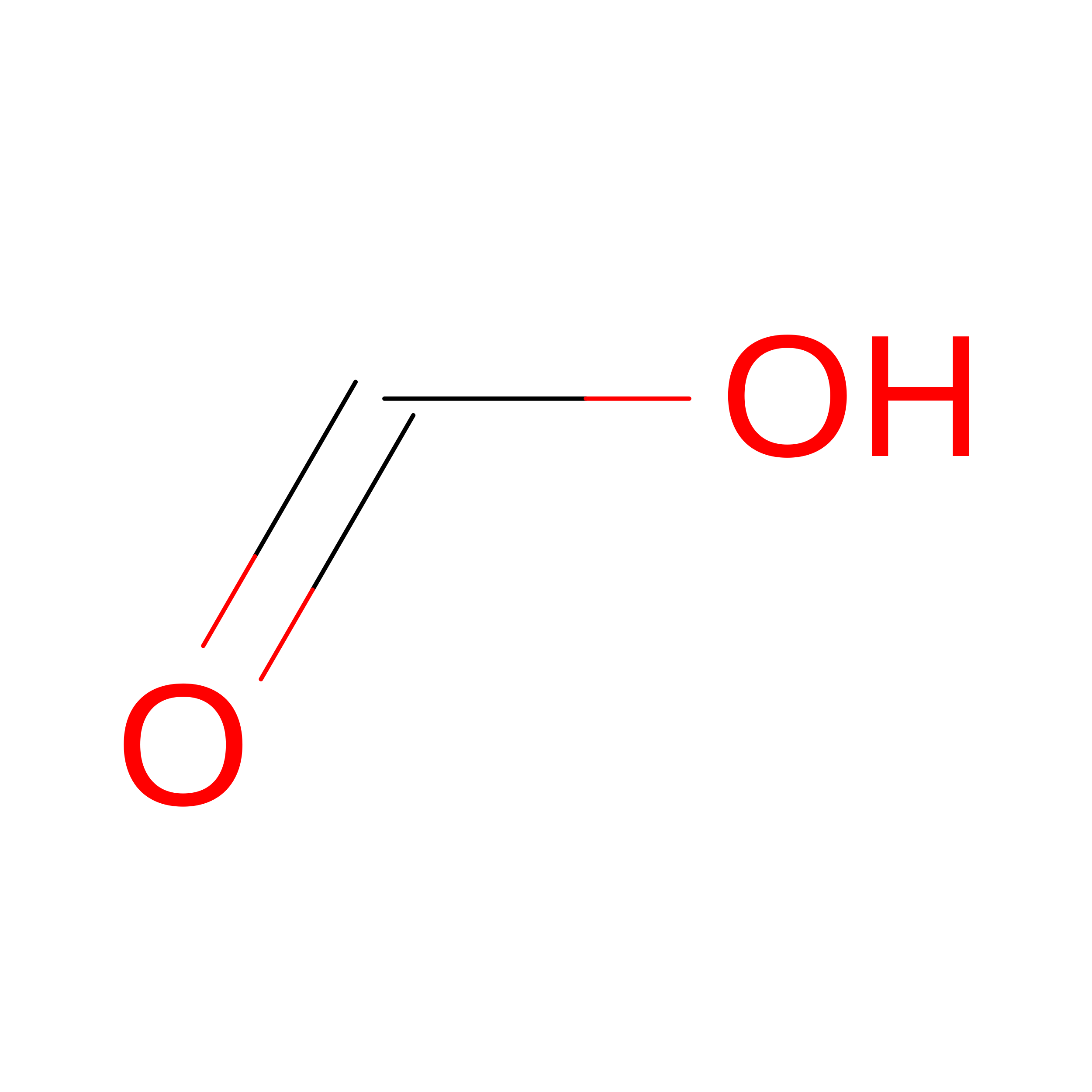
FORMIC ACID
C H2 O2
BDAGIHXWWSANSR-UHFFFAOYSA-N

Experimental Data & Validation

Experimental Data

  • Method: X-RAY DIFFRACTION
  • Resolution: 2.20 Å
  • R-Value Free:  0.234 (Depositor), 0.233 (DCC) 
  • R-Value Work:  0.186 (Depositor), 0.186 (DCC) 
  • R-Value Observed: 0.188 (Depositor) 
Space Group: P 1
Unit Cell:
Length ( Å )Angle ( ˚ )
a = 40.225α = 88.19
b = 42.281β = 64.78
c = 47.088γ = 75.24
Software Package:
Software NamePurpose
PHENIXrefinement
Aimlessdata scaling
XDSdata reduction
MOLREPphasing
PDB_EXTRACTdata extraction

Structure Validation

View Full Validation Report


Entry History 

& Funding Information

Deposition Data


Funding OrganizationLocationGrant Number
Spanish Ministry of Science, Innovation, and UniversitiesSpainPID2023-146281NB-I00

Revision History  (Full details and data files)

  • Version 1.0: 2026-09-02
    Type: Initial release