9SJR | pdb_00009sjr

Cryo-EM structure of Human Apoferritin at pH 3.5


Experimental Data Snapshot

  • Method: ELECTRON MICROSCOPY
  • Resolution: 1.99 Å
  • Aggregation State: PARTICLE 
  • Reconstruction Method: SINGLE PARTICLE 

Starting Model: experimental
View more details

wwPDB Validation   3D Report Full Report


This is version 1.1 of the entry. See complete history


Literature

Direct evidence of acid-driven protein desolvation.

Hamdi, F.Skalidis, I.Schwerin, I.K.Belapure, J.Semchonok, D.A.Kyrilis, F.L.Tuting, C.Muller, J.Kunze, G.Kastritis, P.L.

(2026) Proc Natl Acad Sci U S A 123: e2525949123-e2525949123

  • DOI: https://doi.org/10.1073/pnas.2525949123
  • Primary Citation Related Structures: 
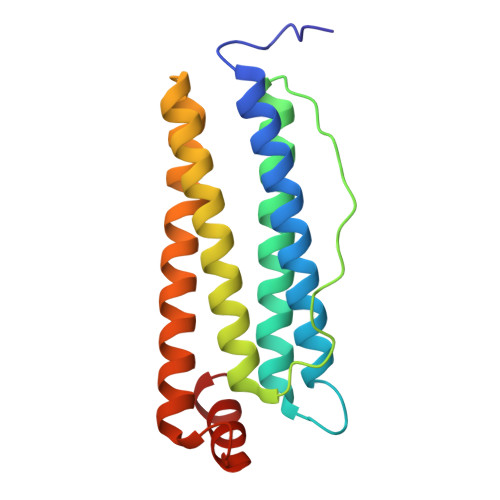
    9SJR, 9SJS, 9SJT, 9SJU, 9SJV

  • PubMed Abstract: 

    Water and its ability to modulate the protonation states of biomolecules govern the physical chemistry of life, dictating their metabolic functions. However, how amino acid protonation alters protein hydration and solubility is an open question since Kuntz and Kauzmann proposed p H -driven protein desolvation in 1974. Here, in a series of high-resolution cryoelectron microscopy structures of a protein complex at different p H values (from p H 9.0 to 3.5), we examined thousands of observable hydration sites. Cryoelectron microscopy data, in agreement with constant-p H molecular dynamics simulations, show that nearly half of protein-bound waters exchanged with the bulk solvent upon acidification, with ~100 waters lost per p H unit per molecule. The loss of waters was most significant around the side chains of glutamate and aspartate residues while specific polar residues, mostly asparagine, anchored persistent waters. A positionally conserved hydration layer was observed across all p H conditions, accounting for 40% of resolved waters. Those waters displayed denser packing than less persistent waters, forming a p H -independent solvation shell. Acid-induced water exchange also displaced bound iron, providing a mechanistic link between solvation and metal release. Our findings demonstrate the core principles of acid-driven protein desolvation, resolving a 50-y-old biochemical hypothesis.


  • Organizational Affiliation
    • Department of Integrative Structural Biochemistry, Institute of Biochemistry and Biotechnology, Martin Luther University Halle-Wittenberg, Halle/Saale 06120, Germany.

Macromolecules
Find similar proteins by:  (by identity cutoff)  |  3D Structure
Entity ID: 1
MoleculeChains Sequence LengthOrganismDetailsImage
Ferritin heavy chain, N-terminally processed173Homo sapiensMutation(s): 0 
Gene Names: FTH1FTHFTHL6OK/SW-cl.84PIG15
EC: 1.16.3.1
UniProt & NIH Common Fund Data Resources
Find proteins for P02794 (Homo sapiens)
Explore P02794 
Go to UniProtKB:  P02794
PHAROS:  P02794
GTEx:  ENSG00000167996 
Entity Groups  
Sequence Clusters30% Identity50% Identity70% Identity90% Identity95% Identity100% Identity
UniProt GroupP02794
Sequence Annotations
Expand
  • Reference Sequence
Small Molecules
Ligands 2 Unique
IDChains Name / Formula / InChI Key2D Diagram3D Interactions
FE
Query on FE

Download Ideal Coordinates CCD File 
AA [auth A]
AB [auth J]
BA [auth A]
BB [auth J]
CA [auth A]
AA [auth A],
AB [auth J],
BA [auth A],
BB [auth J],
CA [auth A],
FA [auth D],
HA [auth D],
IA [auth D],
JA [auth D],
JB [auth R],
LA [auth F],
LB [auth R],
MB [auth R],
NA [auth F],
NB [auth R],
OA [auth F],
PA [auth F],
SA [auth I],
UA [auth I],
VA [auth I],
WA [auth I],
XA [auth J],
Y [auth A],
ZA [auth J]
FE (III) ION
Fe
VTLYFUHAOXGGBS-UHFFFAOYSA-N
MG
Query on MG

Download Ideal Coordinates CCD File 
CB [auth K]
DA [auth B]
DB [auth L]
EA [auth C]
EB [auth M]
CB [auth K],
DA [auth B],
DB [auth L],
EA [auth C],
EB [auth M],
FB [auth N],
GA [auth D],
GB [auth O],
HB [auth P],
IB [auth Q],
KA [auth E],
KB [auth R],
MA [auth F],
OB [auth S],
PB [auth T],
QA [auth G],
QB [auth V],
RA [auth H],
RB [auth W],
SB [auth X],
TA [auth I],
TB [auth Y],
YA [auth J],
Z [auth A]
MAGNESIUM ION
Mg
JLVVSXFLKOJNIY-UHFFFAOYSA-N
Experimental Data & Validation

Experimental Data

  • Method: ELECTRON MICROSCOPY
  • Resolution: 1.99 Å
  • Aggregation State: PARTICLE 
  • Reconstruction Method: SINGLE PARTICLE 
EM Software:
TaskSoftware PackageVersion
RECONSTRUCTIONRELION4
MODEL REFINEMENTPHENIX

Structure Validation

View Full Validation Report


Entry History & Funding Information

Deposition Data


Funding OrganizationLocationGrant Number
European Union (EU)European Union101086665
German Federal Ministry for Education and ResearchGermany03Z22HN23
German Federal Ministry for Education and ResearchGermany03Z22HI2
German Federal Ministry for Education and ResearchGermany03COV04
European Regional Development FundEuropean UnionZS/2016/04/78115
European Regional Development FundEuropean UnionZS/2024/05/187255
German Research Foundation (DFG)Germany391498659
German Research Foundation (DFG)Germany514901783
Fundacao para a Ciencia e a TecnologiaPortugalUIDB/04612/2020
Fundacao para a Ciencia e a TecnologiaPortugalUIDP/04612/2020
Fundacao para a Ciencia e a TecnologiaPortugalLA/P/0087/2020
H2020 Marie Curie Actions of the European CommissionEuropean Union101207412

Revision History  (Full details and data files)

  • Version 1.0: 2026-02-25
    Type: Initial release
  • Version 1.1: 2026-03-18
    Changes: Data collection, Database references