Seeding biosensor cell line that reproduces the Alzheimer tau fold.
Katsinelos, T., Lovestam, S., Qi, C., Ryskeldi-Falcon, B., Macdonald, J.A., Stephenson, G., Ghetti, B., McKee, A., Scheres, S.H.W., Goedert, M.(2025) J Biological Chem 301: 110952-110952
- PubMed: 41260337 Search on PubMed
- DOI: https://doi.org/10.1016/j.jbc.2025.110952
- Primary Citation Related Structures:
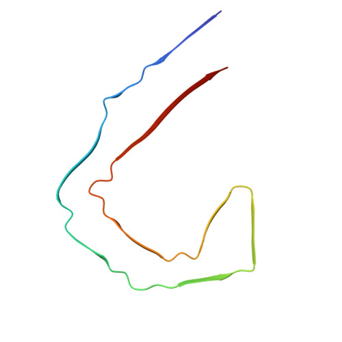
9SHS - PubMed Abstract:
The assembly of tau protein into amyloid filaments through templated seeding is believed to underlie the propagation of pathology in neurodegenerative diseases, such as Alzheimer's disease (AD) and other tauopathies. A commonly used model system for studying this process is through the induction of tau filament formation in cultured cells following the addition of tau seeds isolated from the human brain. However, little is known about the structures of seeded filaments; some biosensor cell lines are unable to reproduce the tau filament structures from AD, because they overexpress tau fragments that do not cover the whole of the ordered filament core. Here, we describe a novel tau seeding biosensor model in HEK293T cells that overexpress residues K297-E391 of human 4R human tau. The construct contains an N-terminal HA-tag, which allows the specific detection of the amplified template. The biosensor cells detected filaments seeded by material from sporadic 3R + 4R tauopathies, with little activity by seeds from 3R-only or 4R-only tauopathies. The sensitivity of seed detection from 3R + 4R tauopathies in our system was similar or higher than for previously reported biosensors. We also structurally characterised the AD-seeded tau filaments by electron cryo-microscopy (cryo-EM). Most of the cell-derived filaments consisted of two protofilaments with the Alzheimer fold, but with a 'head-to-head' inter-protofilament packing. Our results establish a sensitive biosensor cell line with specificity towards seeds from 3R + 4R tauopathies.
- MRC Laboratory of Molecular Biology, Cambridge, UK.
Organizational Affiliation: