9SHQ | pdb_00009shq

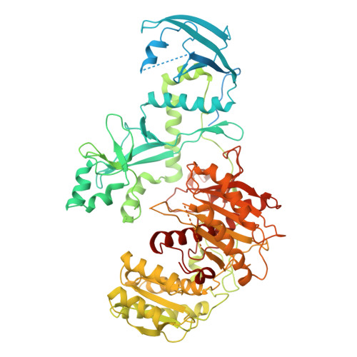
Cryo-EM structure of the endogeneous MIWI in complex with pachytene piRNA at 4.2A


Experimental Data Snapshot

  • Method: ELECTRON MICROSCOPY
  • Resolution: 4.20 Å
  • Aggregation State: PARTICLE 
  • Reconstruction Method: SINGLE PARTICLE 

wwPDB Validation 3D Report Full Report

Validation slider image for 9SHQ

This is version 1.0 of the entry. See complete history

Literature

Structure of the MIWI endoribonuclease bound to pachytene piRNAs from mouse testes.

Raad, N.Fernandez-Rodriguez, C.Pandey, R.R.Mohammed, I.Uchikawa, E.Burger, F.Homolka, D.Pillai, R.S.

(2026) Cell Rep 45: 116804-116804

  • DOI: https://doi.org/10.1016/j.celrep.2025.116804
  • Primary Citation Related Structures: 
    9S0Z, 9S1E, 9SHP, 9SHQ

  • PubMed Abstract: 

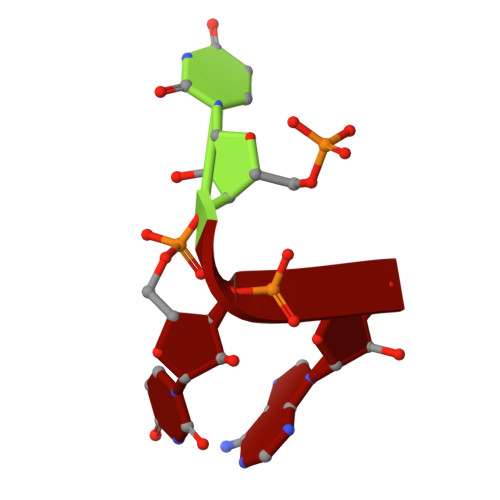
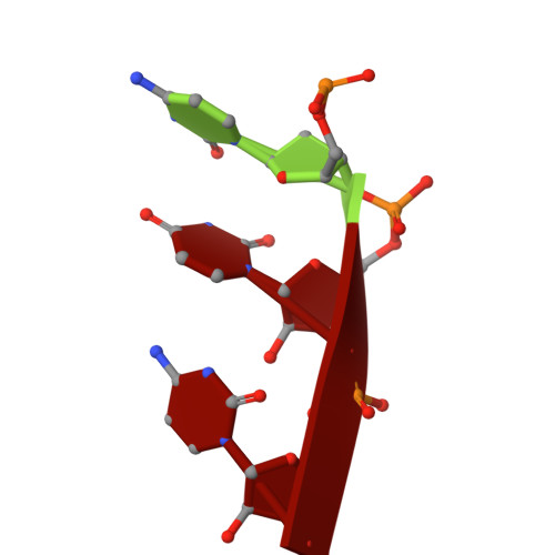
    PIWI-interacting RNAs (piRNAs) guide PIWI endoribonucleases to destroy transposon transcripts, ensuring animal fertility. Here, we report the cryo-electron microscopy structure of the MIWI-pachytene piRNA complex isolated from mouse testes. The piRNA is held via non-specific charge-based interactions with the RNA backbone and by specific recognition of the first nucleotide uridine by residues within the MID and PIWI domains. The first six nucleotides of the guide RNA take up the A-form conformation to facilitate pairing with the target. The RNA channel is wider than that observed in insect PIWI proteins, explaining the tolerance for piRNA seed:target mismatches. The PIWI endonuclease domain is in an inactive "un-plugged" state, with the loop containing a catalytic residue (E671) requiring structural re-orientation for activity. Furthermore, the PIWI domain reveals a conserved pre-formed pocket that may serve to accommodate a conserved tryptophan from the interacting factor GTSF1 to promote small RNA-guided endoribonuclease activity.


  • Organizational Affiliation
    • Department of Molecular Biology, Science III, University of Geneva, 30 Quai Ernest-Ansermet, Geneva 1211, Switzerland. Electronic address: nicole.raad@unige.ch.

Macromolecule Content 

  • Total Structure Weight: 101.54 kDa 
  • Atom Count: 5,409 
  • Modeled Residue Count: 667 
  • Deposited Residue Count: 876 
  • Unique protein chains: 1
  • Unique nucleic acid chains: 2

Macromolecules


Find similar proteins by:|  3D Structure
Entity ID: 1
MoleculeChains  Sequence LengthOrganismDetailsImage
Piwi-like protein 1A [auth B]870Mus musculusMutation(s): 0 
EC: 3.1.26
UniProt
Find proteins for Q9JMB7 (Mus musculus)
Explore Q9JMB7 
Go to UniProtKB:  Q9JMB7
Entity Groups
Sequence Clusters30% Identity50% Identity70% Identity90% Identity95% Identity100% Identity
UniProt GroupQ9JMB7
Sequence Annotations
Expand
Reference Sequence
Find similar nucleic acids by:  Sequence
Entity ID: 2
MoleculeChains LengthOrganismImage
RNA (5'-R(P*UP*UP*A)-3')B [auth C]3Mus musculus
Sequence Annotations
Expand
Reference Sequence
Find similar nucleic acids by:  Sequence
Entity ID: 3
MoleculeChains LengthOrganismImage
RNA (5'-R(P*CP*UP*C)-3')C [auth D]3Mus musculus
Sequence Annotations
Expand
Reference Sequence

Small Molecules

Ligands 1 Unique
IDChains Name / Formula / InChI Key2D Diagram3D Interactions
MG
(Subject of Investigation/LOI)

Query on MG


Download:Ideal Coordinates CCD File
D [auth B]MAGNESIUM ION
Mg
JLVVSXFLKOJNIY-UHFFFAOYSA-N

Experimental Data & Validation

Experimental Data

  • Method: ELECTRON MICROSCOPY
  • Resolution: 4.20 Å
  • Aggregation State: PARTICLE 
  • Reconstruction Method: SINGLE PARTICLE 
EM Software:
TaskSoftware PackageVersion
MODEL REFINEMENTPHENIX1.20.1_4487
RECONSTRUCTIONcryoSPARC

Structure Validation

View Full Validation Report


Entry History 

& Funding Information

Deposition Data


Funding OrganizationLocationGrant Number
Swiss National Science FoundationSwitzerland310030_215346
Swiss National Science FoundationSwitzerland#51NF40-205601
Other privateThe Lalor Foundation

Revision History  (Full details and data files)

  • Version 1.0: 2026-01-21
    Type: Initial release