9SGK | pdb_00009sgk

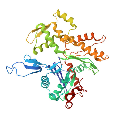
F-actin in complex with USP54 M1 actin binding motif


Experimental Data Snapshot

  • Method: ELECTRON MICROSCOPY
  • Resolution: 3.80 Å
  • Aggregation State: FILAMENT 
  • Reconstruction Method: HELICAL 

wwPDB Validation 3D Report Full Report

Validation slider image for 9SGK

This is version 1.1 of the entry. See complete history

Literature

Evolutionarily conserved short linear motifs drive actin filament binding.

Paraschiakos, T.Yuan, B.Hecht-Bucher, M.Sopelniak, K.Cervero, P.Simon, L.Zonjic, K.Eggers, D.Selle, F.Li, J.Biabani, A.Linder, S.Marlovits, T.C.Windhorst, S.

(2026) Nat Cell Biol 28: 1437-1452

  • DOI: https://doi.org/10.1038/s41556-026-01979-9
  • Primary Citation Related Structures: 
    8R3H, 9QGK, 9SGK

  • PubMed Abstract: 

    Regulation of the actin cytoskeleton by actin-binding proteins is essential for cellular homeostasis, and the mode of actin binding determines the activity of actin-binding proteins. Here we identify a 'short linear actin filament-binding motif' (SFM) based on the cryo-electron microscopy structure of the ITPKA-actin filament complex. Using the computational pipeline SLiMFold, we discovered 103 human proteins containing SFMs with diverse cellular roles. Phylogenetic analysis suggests that SFMs arose de novo and are conserved across eukaryotes, exhibiting actin filament-binding affinities of 2-12 µM. Critical residues mediating binding and modulating affinity were defined, and the cryo-electron microscopy structures of two SFM-actin filament complexes revealed that SFM binding decreases actin-filament stiffness. These findings indicate that SFMs regulate actin-filament conformation and serve as anchoring modules that connect actin dynamics to a broad variety of cellular functions, providing a framework for understanding the actin-associated roles of numerous proteins.


  • Organizational Affiliation
    • Department of Biochemistry and Signal Transduction, University Medical Center Hamburg-Eppendorf, Hamburg, Germany.

Macromolecule Content 

  • Total Structure Weight: 380.45 kDa 
  • Atom Count: 15,200 
  • Modeled Residue Count: 1,920 
  • Deposited Residue Count: 3,390 
  • Unique protein chains: 2

Macromolecules

Find similar proteins by:|  3D Structure
Entity ID: 1
MoleculeChains  Sequence LengthOrganismDetailsImage
Actin binding motifA [auth B],
B [auth C],
C [auth D],
D [auth E],
J [auth P]
301Homo sapiensMutation(s): 0 
Entity Groups
Sequence Clusters30% Identity50% Identity70% Identity90% Identity95% Identity100% Identity
Sequence Annotations
Expand
Reference Sequence
Find similar proteins by:|  3D Structure
Entity ID: 2
MoleculeChains  Sequence LengthOrganismDetailsImage
Actin, alpha skeletal muscleE [auth F],
F [auth G],
G [auth H],
H [auth I],
I [auth J]
377Gallus gallusMutation(s): 0 
EC: 3.6.4
UniProt
Find proteins for P68139 (Gallus gallus)
Explore P68139 
Go to UniProtKB:  P68139
Entity Groups
Sequence Clusters30% Identity50% Identity70% Identity90% Identity95% Identity100% Identity
UniProt GroupP68139
Sequence Annotations
Expand
Reference Sequence

Small Molecules

Ligands 2 Unique
IDChains Name / Formula / InChI Key2D Diagram3D Interactions
ADP
(Subject of Investigation/LOI)

Query on ADP


Download:Ideal Coordinates CCD File
L [auth F],
N [auth G],
P [auth H],
R [auth I],
T [auth J]
ADENOSINE-5'-DIPHOSPHATE
C10 H15 N5 O10 P2
XTWYTFMLZFPYCI-KQYNXXCUSA-N
MG
(Subject of Investigation/LOI)

Query on MG


Download:Ideal Coordinates CCD File
K [auth F],
M [auth G],
O [auth H],
Q [auth I],
S [auth J]
MAGNESIUM ION
Mg
JLVVSXFLKOJNIY-UHFFFAOYSA-N
Modified Residues  1 Unique
IDChains TypeFormula2D DiagramParent
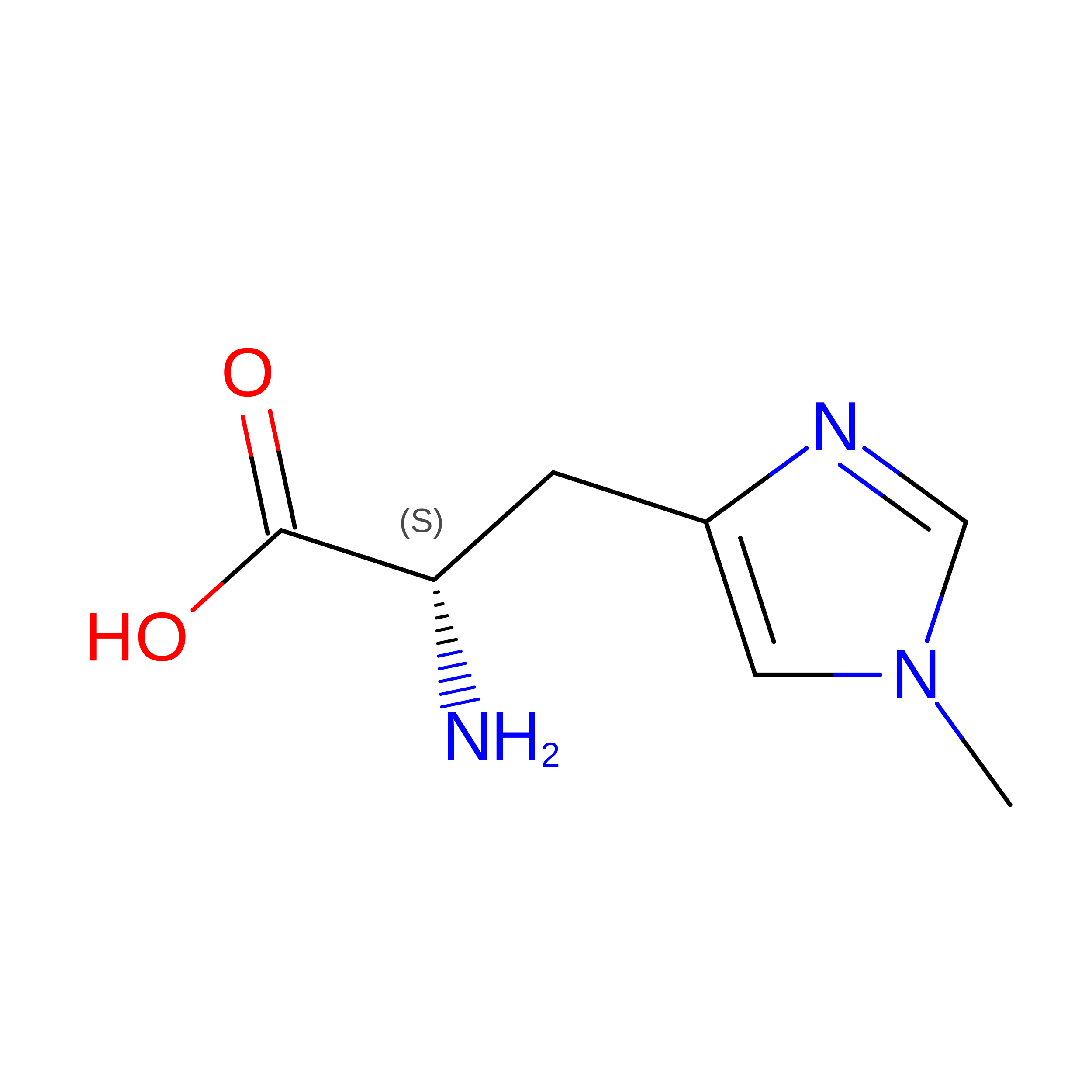
HIC
Query on HIC
E [auth F],
F [auth G],
G [auth H],
H [auth I],
I [auth J]
L-PEPTIDE LINKINGC7 H11 N3 O2HIS

Experimental Data & Validation

Experimental Data

  • Method: ELECTRON MICROSCOPY
  • Resolution: 3.80 Å
  • Aggregation State: FILAMENT 
  • Reconstruction Method: HELICAL 
EM Software:
TaskSoftware PackageVersion
RECONSTRUCTIONcryoSPARC

Structure Validation

View Full Validation Report


Entry History 

& Funding Information

Deposition Data


Funding OrganizationLocationGrant Number
Not funded--

Revision History  (Full details and data files)

  • Version 1.0: 2026-04-29
    Type: Initial release
  • Version 1.1: 2026-07-29
    Changes: Data collection, Database references