9SDY | pdb_00009sdy

Structure of RBR E2 variant binding to CUL5-RBX2 bound ARIH2


Experimental Data Snapshot

  • Method: ELECTRON MICROSCOPY
  • Resolution: 3.06 Å
  • Aggregation State: PARTICLE 
  • Reconstruction Method: SINGLE PARTICLE 

wwPDB Validation   3D Report Full Report


This is version 1.1 of the entry. See complete history


Literature

E2 variants for probing E3 ubiquitin ligase activities.

Du, J.Andree, G.A.Horn-Ghetko, D.Stier, L.Singh, J.Kostrhon, S.Kiss, L.Mann, M.Sidhu, S.S.Schulman, B.A.

(2026) Proc Natl Acad Sci U S A 123: e2524899122-e2524899122

  • DOI: https://doi.org/10.1073/pnas.2524899122
  • Primary Citation of Related Structures:  
    9SDX, 9SDY

  • PubMed Abstract: 

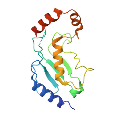
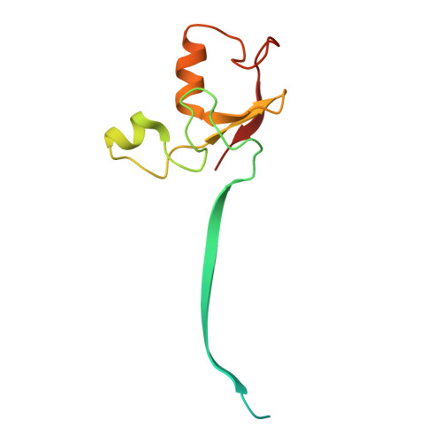
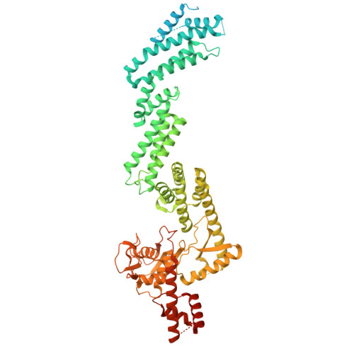
    E3 ligases partner with E2 enzymes to regulate vast eukaryotic biology. The hierarchical nature of these pairings, with >600 E3s and ~40 E2s in humans, necessitates that E2s cofunction with numerous different E3s. Here, focusing on E3s in the RING-between-RING (RBR) family and their partner UBE2L3 and UBE2D-family E2s, we report an approach to interrogate selected pathways. We screened phage-displayed libraries of structure-based E2 variants (E2Vs) to discover enzymes with enhanced affinity and specificity toward half of all RBR E3 ligases (ARIH1, ARIH2, ANKIB1, CUL9, HOIL1, HOIP, and RNF14). Collectively, these E2Vs allowed distinguishing actions of different cofunctioning E3s, obtaining high-resolution cryogenic Electron Microscopy (cryo-EM) structures of an RBR E3 in the context of a substrate-bound multiprotein complex, and profiling an endogenous RBR E3 response to an extracellular stimulus. Overall, we anticipate that E2V technology will be a generalizable tool to enable in-depth mechanistic and structural analysis of E3 ligase functions, and mapping their activity states and protein partners in cellular signaling cascades.


  • Organizational Affiliation
    • Department of Molecular Machines and Signaling, Max Planck Institute of Biochemistry, Martinsried 82152, Germany.

Macromolecules
Find similar proteins by:  (by identity cutoff)  |  3D Structure
Entity ID: 1
MoleculeChains Sequence LengthOrganismDetailsImage
L3A2-1A [auth G]155synthetic constructMutation(s): 0 
Entity Groups  
Sequence Clusters30% Identity50% Identity70% Identity90% Identity95% Identity100% Identity
Sequence Annotations
Expand
  • Reference Sequence
Find similar proteins by:  (by identity cutoff)  |  3D Structure
Entity ID: 2
MoleculeChains Sequence LengthOrganismDetailsImage
RING-box protein 2B [auth R]113Homo sapiensMutation(s): 0 
Gene Names: RNF7RBX2ROC2SAG
EC: 2.3.2.27 (PDB Primary Data), 2.3.2.32 (PDB Primary Data)
UniProt & NIH Common Fund Data Resources
Find proteins for Q9UBF6 (Homo sapiens)
Explore Q9UBF6 
Go to UniProtKB:  Q9UBF6
PHAROS:  Q9UBF6
GTEx:  ENSG00000114125 
Entity Groups  
Sequence Clusters30% Identity50% Identity70% Identity90% Identity95% Identity100% Identity
UniProt GroupQ9UBF6
Sequence Annotations
Expand
  • Reference Sequence
Find similar proteins by:  (by identity cutoff)  |  3D Structure
Entity ID: 3
MoleculeChains Sequence LengthOrganismDetailsImage
Cullin-5780Homo sapiensMutation(s): 0 
Gene Names: CUL5VACM1
UniProt & NIH Common Fund Data Resources
Find proteins for Q93034 (Homo sapiens)
Explore Q93034 
Go to UniProtKB:  Q93034
PHAROS:  Q93034
GTEx:  ENSG00000166266 
Entity Groups  
Sequence Clusters30% Identity50% Identity70% Identity90% Identity95% Identity100% Identity
UniProt GroupQ93034
Sequence Annotations
Expand
  • Reference Sequence
Find similar proteins by:  (by identity cutoff)  |  3D Structure
Entity ID: 4
MoleculeChains Sequence LengthOrganismDetailsImage
E3 ubiquitin-protein ligase ARIH2D [auth H]493Homo sapiensMutation(s): 0 
Gene Names: ARIH2ARI2TRIAD1HT005
EC: 2.3.2.31
UniProt & NIH Common Fund Data Resources
Find proteins for O95376 (Homo sapiens)
Explore O95376 
Go to UniProtKB:  O95376
PHAROS:  O95376
GTEx:  ENSG00000177479 
Entity Groups  
Sequence Clusters30% Identity50% Identity70% Identity90% Identity95% Identity100% Identity
UniProt GroupO95376
Sequence Annotations
Expand
  • Reference Sequence
Experimental Data & Validation

Experimental Data

  • Method: ELECTRON MICROSCOPY
  • Resolution: 3.06 Å
  • Aggregation State: PARTICLE 
  • Reconstruction Method: SINGLE PARTICLE 
EM Software:
TaskSoftware PackageVersion
MODEL REFINEMENTPHENIX1.21.1_5286
RECONSTRUCTIONcryoSPARC

Structure Validation

View Full Validation Report


Entry History & Funding Information

Deposition Data


Funding OrganizationLocationGrant Number
German Research Foundation (DFG)GermanySCHU3196/1-1

Revision History  (Full details and data files)

  • Version 1.0: 2025-12-24
    Type: Initial release
  • Version 1.1: 2026-01-14
    Changes: Data collection, Database references