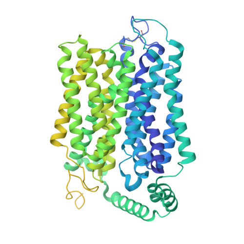
9SDL | pdb_00009sdl

Cryo-EM structure of PfHT1 bound to 2,5-anhydro-D-mannitol


Experimental Data Snapshot

  • Method: ELECTRON MICROSCOPY
  • Resolution: 2.42 Å
  • Aggregation State: PARTICLE 
  • Reconstruction Method: SINGLE PARTICLE 

wwPDB Validation 3D Report Full Report

Validation slider image for 9SDL

This is version 1.2 of the entry. See complete history

Literature

A two-step mechanism for sugar translocation.

Ahn, D.H.Alleva, C.Reichenbach, T.Gulati, A.Ruda, A.Bonaccorsi, M.Silberberg, J.M.Claesson, M.Suades, A.Delemotte, L.Widmalm, G.Drew, D.

(2026) Nat Struct Mol Biol 33: 652-663

  • DOI: https://doi.org/10.1038/s41594-026-01784-w
  • Primary Citation Related Structures: 
    9SDL

  • PubMed Abstract: 

    In mammals, glucose transporters (GLUTs) mediate organism-wide sugar distribution, yet the molecular basis of substrate specificity remains unclear. The bacterial xylose transporter XylE serves as a model for GLUTs. However, although xylose and glucose bind with a similar affinity, xylose is transported, but glucose acts as an inhibitor. Here, using saturation transfer difference (STD) nuclear magnetic resonance (NMR) spectroscopy, we distinguished transported sugars from sugar inhibitors. Our findings revealed that only transported sugars generate STD NMR signals, which are abolished for xylose when XylE is trapped in either outward- or inward-facing conformations. Engineering the sugar-binding pocket and gating helix TM7b enabled glucose transport by XylE and corresponding STD signals. Using complementary molecular dynamics simulations, together with structural, biochemical and STD NMR analysis of related parasitic and mammalian GLUTs, we identified TM7b as a key determinant of occluded state formation. We conclude that, rather than the initial substrate-binding event observed in experimental structures, formation of a substrate-induced transition-state intermediate is the primary determinant of specificity in transporters.


  • Organizational Affiliation
    • Department of Biochemistry and Biophysics, Science for Life Laboratory, Stockholm University, Stockholm, Sweden.

Macromolecule Content 

  • Total Structure Weight: 169.61 kDa 
  • Atom Count: 7,604 
  • Modeled Residue Count: 956 
  • Deposited Residue Count: 1,498 
  • Unique protein chains: 1

Macromolecules

Find similar proteins by:|  3D Structure
Entity ID: 1
MoleculeChains  Sequence LengthOrganismDetailsImage
Hexose transporter 1,Green fluorescent protein
A, B
749Plasmodium falciparumAequorea victoria
This entity is chimeric
Mutation(s): 0 
Gene Names: ht1GFP
UniProt
Find proteins for O97467 (Plasmodium falciparum)
Explore O97467 
Go to UniProtKB:  O97467
Find proteins for P42212 (Aequorea victoria)
Explore P42212 
Go to UniProtKB:  P42212
Entity Groups
Sequence Clusters30% Identity50% Identity70% Identity90% Identity95% Identity100% Identity
UniProt GroupsP42212O97467
Sequence Annotations
Expand
Reference Sequence

Experimental Data & Validation

Experimental Data

  • Method: ELECTRON MICROSCOPY
  • Resolution: 2.42 Å
  • Aggregation State: PARTICLE 
  • Reconstruction Method: SINGLE PARTICLE 
EM Software:
TaskSoftware PackageVersion
RECONSTRUCTIONcryoSPARC

Structure Validation

View Full Validation Report


Entry History 

& Funding Information

Deposition Data


Funding OrganizationLocationGrant Number
Knut and Alice Wallenberg FoundationSweden--
Novo Nordisk FoundationDenmark--
CancerfondenSweden--

Revision History  (Full details and data files)

  • Version 1.0: 2026-04-15
    Type: Initial release
  • Version 1.1: 2026-04-22
    Changes: Data collection, Database references
  • Version 1.2: 2026-04-29
    Changes: Data collection, Database references