Pushing the limits of hydrogen/deuterium exchange mass spectrometry to study protein:fragment low affinity interactions.
F Malta, C., O Silva, D., Gradler, U., M F Sousa, P., Musil, D., Schwarz, D., Bomke, J., M Bandeiras, T., Bortoluzzi, A.(2025) Commun Chem 8: 405-405
- PubMed: 41429916 Search on PubMedSearch on PubMed Central
- DOI: https://doi.org/10.1038/s42004-025-01787-6
- Primary Citation Related Structures:
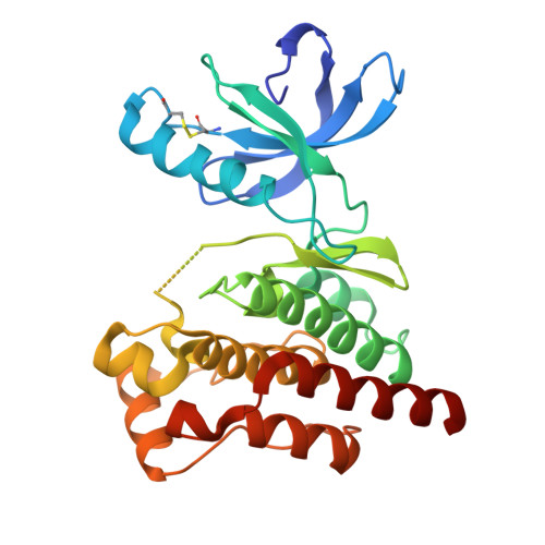
9SDI - PubMed Abstract:
Characterization of protein-ligand interactions is essential for the pre-clinical development of drug candidates and Hydrogen/Deuterium Exchange Mass Spectrometry (HDX-MS) has emerged as a valuable tool in this process. HDX-MS has predominantly been employed with high affinity compounds with only a few examples of its application for weaker binders such as fragments. Nevertheless, HDX-MS usage could be instrumental in Fragment-Based Drug Discovery (FBDD) programs. In this work, the drug-target protein Cyclophilin D (CypD) was used as a model to explore the boundaries of fragments binding characterization by HDX-MS (fHDX-MS). We performed a systematic study on the optimal conditions for fHDX-MS execution and found that fragments with binding affinities in the double-digit mM range are still amenable to fHDX-MS. We observed that, despite the intrinsic low resolution of HDX-MS, fragments binding sites that partially overlap can still be distinguished. Overall, this study shows that fHDX-MS can be a useful method for FBDD.
- iBET-Instituto de Biologia Experimental e Tecnológica, Av. da República, Oeiras, Portugal.
Organizational Affiliation: