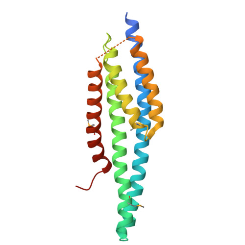
Molecular structure of the ESCRT-III-based archaeal CdvAB cell division machinery.
Drobnic, T., Salzer, R., Nierhaus, T., Jiang, M.K.X., Bellini, D., Steindorf, A., Albers, S.V., Baum, B., Lowe, J.(2026) Proc Natl Acad Sci U S A 123: e2525941123-e2525941123
- PubMed: 41543908 Search on PubMedSearch on PubMed Central
- DOI: https://doi.org/10.1073/pnas.2525941123
- Primary Citation Related Structures:
9S97, 9S98, 9S99, 9S9G, 9S9H, 9S9I, 9S9J, 9S9K - PubMed Abstract:
Most prokaryotes divide using filaments of the tubulin-like FtsZ protein, while some archaea employ instead ESCRT-III-like proteins and their filaments for cell division and cytokinesis. The alternative archaeal system comprises Cdv proteins and is thought to bear some resemblance to ESCRT-III-based membrane remodeling in other domains of life, including eukaryotes, especially during abscission. Here, we present biochemical, crystallographic, and cryo-EM studies of the Sulfolobus Cdv machinery. CdvA, an early non-ESCRT component, adopts a PRC-domain/coiled-coil fold and polymerizes into long double-stranded helical filaments, mainly via hydrophobic interfaces. Monomeric CdvB adopts the canonical ESCRT-III fold in both a closed and a distinct "semiopen" conformation. Soluble CdvB2 filaments are composed of subunits in the closed state, appearing to transition to the open, active state only when polymerized on membranes. Short N-terminal amphipathic helices in all CdvB paralogues, B, B1, and B2, mediate membrane binding and are required for liposome recruitment in vitro. We provide a molecular overview of archaeal ESCRT-III-based cytokinesis machinery, the definitive demonstration that CdvB proteins are bona fide ESCRT-III homologues, and reveal the molecular basis for membrane engagement. Thus, we illuminate conserved principles of ESCRT-mediated membrane remodeling and extend them to an anciently diverged archaeal lineage.
- Medical Research Council Laboratory of Molecular Biology, Structural Studies Division, Cambridge CB2 0QH, United Kingdom.
Organizational Affiliation: