9S60 | pdb_00009s60

Structural and proteomics analysis of the mouse cathepsin B - DARPin 4m3 complex reveals determinants of species - specific binding


Experimental Data Snapshot

  • Method: X-RAY DIFFRACTION
  • Resolution: 1.69 Å
  • R-Value Free: 
    0.216 (Depositor), 0.197 (DCC) 
  • R-Value Work: 
    0.193 (Depositor), 0.197 (DCC) 
  • R-Value Observed: 
    0.193 (Depositor) 

Starting Model: experimental
View more details

wwPDB Validation 3D Report Full Report

Validation slider image for 9S60

This is version 1.0 of the entry. See complete history

Literature

Structural and Proteomic Analysis of the Mouse Cathepsin B-DARPin 4m3 Complex Reveals Species-Specific Binding Determinants.

Zaric, M.Tusar, L.Kramer, L.Vasiljeva, O.Novak, M.Impens, F.Usenik, A.Gevaert, K.Turk, D.Turk, B.

(2025) Int J Mol Sci 26

  • DOI: https://doi.org/10.3390/ijms262411910
  • Primary Citation Related Structures: 
    9S60

  • PubMed Abstract: 

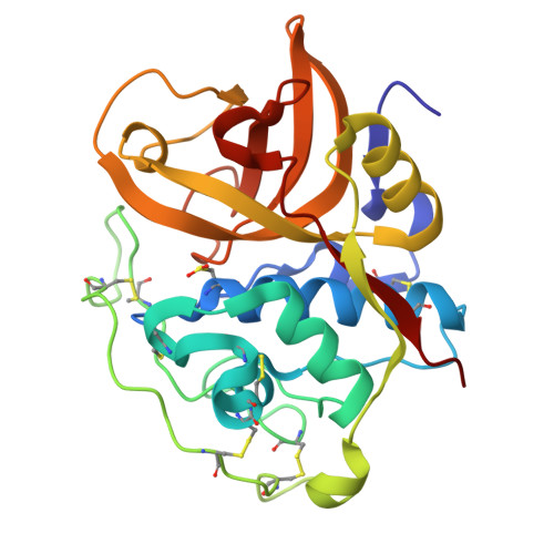
    Cathepsin B (CatB) is a lysosomal cysteine protease that plays a major role in various pathologies and is therefore considered a valuable therapeutic target. To address species-specific inhibitor challenges, we characterized the selective binding of designed ankyrin repeat protein (DARPin) 4m3 toward mouse cathepsin B (mCatB) over human CatB (hCatB). The mCatB-DARPin 4m3 complex was validated by size-exclusion chromatography (SEC), nano-differential scanning fluorimetry (nano-DSF), and surface plasmon resonance (SPR), revealing high affinity binding (K D = 65.7 nM) and potent inhibition (Ki = 26.7 nM; mixed competitive/noncompetitive). DARPin 4m3 showed no binding/inhibition toward hCatB. The 1.67 Å crystal structure of the complex-the first for mCatB-identified key interaction residues (e.g., I65/Q66 in mCatB vs. S65/M66 in hCatB) conferring selectivity. Proteomic analysis of endogenous substrates using a support vector machine (SVM) revealed greater similarity between mCatB and hCatB cleavages (Area Under the Curve (AUC) = 0.733) than between mCatB and other human cathepsins (AUC = 0.939-0.965). Clustering and SVM methods offer broadly applicable tools for protease specificity profiling in drug discovery. This study demonstrates the utility of DARPins for species-selective targeting and highlights the importance of integrated structural and proteomic approaches for dissecting protein-protein interactions.


  • Organizational Affiliation
    • Molecular and Structural Biology, Department of Biochemistry, Jožef Stefan Institute, SI-1000 Ljubljana, Slovenia.

Macromolecule Content 

  • Total Structure Weight: 92.14 kDa 
  • Atom Count: 7,072 
  • Modeled Residue Count: 826 
  • Deposited Residue Count: 862 
  • Unique protein chains: 2

Macromolecules

Find similar proteins by:|  3D Structure
Entity ID: 1
MoleculeChains  Sequence LengthOrganismDetailsImage
Cathepsin BA,
B [auth A2]
254Mus musculusMutation(s): 0 
Gene Names: Ctsb
EC: 3.4.22.1
UniProt
Find proteins for P10605 (Mus musculus)
Explore P10605 
Go to UniProtKB:  P10605
Entity Groups
Sequence Clusters30% Identity50% Identity70% Identity90% Identity95% Identity100% Identity
UniProt GroupP10605
Sequence Annotations
Expand
Reference Sequence
Find similar proteins by:|  3D Structure
Entity ID: 2
MoleculeChains  Sequence LengthOrganismDetailsImage
DARPin 4m3C [auth B],
D [auth B2]
177synthetic constructMutation(s): 0 

Small Molecules

Modified Residues  1 Unique
IDChains TypeFormula2D DiagramParent
CSO
Query on CSO
A,
B [auth A2]
L-PEPTIDE LINKINGC3 H7 N O3 SCYS

Experimental Data & Validation

Experimental Data

  • Method: X-RAY DIFFRACTION
  • Resolution: 1.69 Å
  • R-Value Free:  0.216 (Depositor), 0.197 (DCC) 
  • R-Value Work:  0.193 (Depositor), 0.197 (DCC) 
  • R-Value Observed: 0.193 (Depositor) 
Space Group: P 21 21 21
Unit Cell:
Length ( Å )Angle ( ˚ )
a = 52.64α = 90
b = 109.92β = 90
c = 142.64γ = 90
Software Package:
Software NamePurpose
MAINrefinement
XDSdata reduction
PHASERphasing

Structure Validation

View Full Validation Report


Entry History 

& Funding Information

Deposition Data


Funding OrganizationLocationGrant Number
Slovenian Research AgencySloveniaP1-0140
Slovenian Research AgencySloveniaP1-0048
Slovenian Research AgencySloveniaIO-0048

Revision History  (Full details and data files)

  • Version 1.0: 2026-01-14
    Type: Initial release