9S41 | pdb_00009s41

Cerebellar GluA1/4 TMD with TARP gamma 7 (focused refinement)


Experimental Data Snapshot

  • Method: ELECTRON MICROSCOPY
  • Resolution: 3.66 Å
  • Aggregation State: PARTICLE 
  • Reconstruction Method: SINGLE PARTICLE 

Starting Models: experimental, in silico
View more details

wwPDB Validation   3D Report Full Report


This is version 1.0 of the entry. See complete history


Literature

Structure and organization of AMPA receptor-TARP complexes in the mammalian cerebellum.

Scrutton, A.M.Sengupta, N.Ivica, J.Stockwell, I.Peak-Chew, S.Singh, B.Suzuki, K.Chang, V.T.McLaughlin, S.H.Krieger, J.M.Aricescu, A.R.Greger, I.H.

(2025) Science : eaeb3577-eaeb3577

  • DOI: https://doi.org/10.1126/science.aeb3577
  • Primary Citation of Related Structures:  
    9S3O, 9S41

  • PubMed Abstract: 

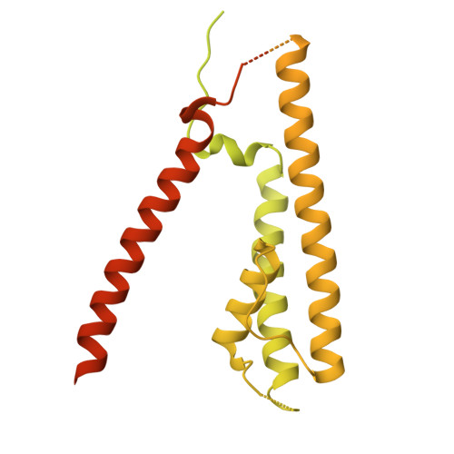
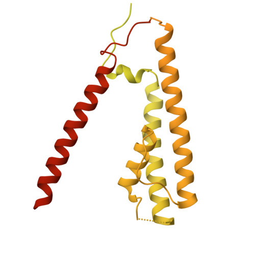
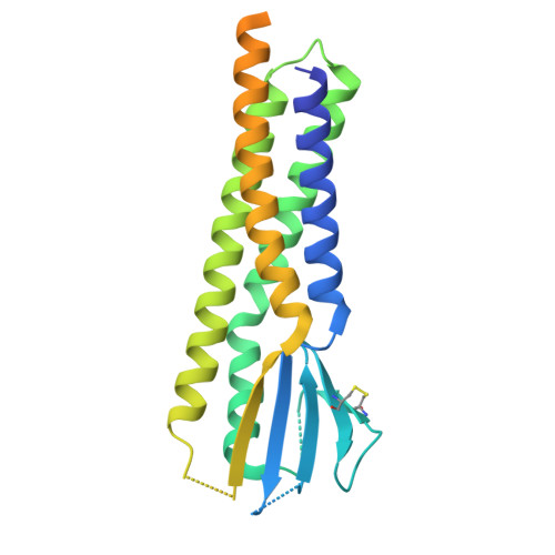
    AMPA receptors (AMPARs) are multimodal transducers of glutamatergic signals throughout the brain. Their diversity is exemplified in the cerebellum; at afferent synapses, AMPARs mediate high-frequency excitation, whereas in Bergmann glia (BG) they support calcium transients that modulate synaptic transmission. This spectrum arises from different combinations of core subunits (GluA1-4), auxiliary proteins, and post-transcriptional modifications. Here, using mass-spectrometry, cryo-EM, and electrophysiology, we characterize major cerebellar AMPARs in pig: calcium-impermeable GluA2/A4 heteromers with four TARP subunits, mainly neuronal in origin, and BG-specific calcium-permeable GluA1/A4 heteromers containing two Type-2 TARPs. We also showed that GluA4 receptors consistently exhibit compact N-terminal domains that promote their synaptic delivery. Our study defines the organizational principles of mammalian cerebellar AMPAR complexes and reveals how different receptor subtypes support cell-type specific functions.


  • Organizational Affiliation
    • Neurobiology Division, Medical Research Council (MRC) Laboratory of Molecular Biology, Cambridge, UK.

Macromolecules
Find similar proteins by:  (by identity cutoff)  |  3D Structure
Entity ID: 1
MoleculeChains Sequence LengthOrganismDetailsImage
Glutamate receptor 1
A, C
888Sus scrofaMutation(s): 0 
UniProt
Find proteins for A0A286ZS63 (Sus scrofa)
Explore A0A286ZS63 
Go to UniProtKB:  A0A286ZS63
Entity Groups  
Sequence Clusters30% Identity50% Identity70% Identity90% Identity95% Identity100% Identity
UniProt GroupA0A286ZS63
Sequence Annotations
Expand
  • Reference Sequence
Find similar proteins by:  (by identity cutoff)  |  3D Structure
Entity ID: 2
MoleculeChains Sequence LengthOrganismDetailsImage
Glutamate receptor 4
B, D
863Sus scrofaMutation(s): 0 
UniProt
Find proteins for I3L8N9 (Sus scrofa)
Explore I3L8N9 
Go to UniProtKB:  I3L8N9
Entity Groups  
Sequence Clusters30% Identity50% Identity70% Identity90% Identity95% Identity100% Identity
UniProt GroupI3L8N9
Sequence Annotations
Expand
  • Reference Sequence
Find similar proteins by:  (by identity cutoff)  |  3D Structure
Entity ID: 3
MoleculeChains Sequence LengthOrganismDetailsImage
Voltage-dependent calcium channel gamma-7 subunitE [auth I],
F [auth J]
275Sus scrofaMutation(s): 0 
UniProt
Find proteins for F2Z4Y2 (Sus scrofa)
Explore F2Z4Y2 
Go to UniProtKB:  F2Z4Y2
Entity Groups  
Sequence Clusters30% Identity50% Identity70% Identity90% Identity95% Identity100% Identity
UniProt GroupF2Z4Y2
Sequence Annotations
Expand
  • Reference Sequence
Experimental Data & Validation

Experimental Data

  • Method: ELECTRON MICROSCOPY
  • Resolution: 3.66 Å
  • Aggregation State: PARTICLE 
  • Reconstruction Method: SINGLE PARTICLE 
EM Software:
TaskSoftware PackageVersion
RECONSTRUCTIONcryoSPARC
MODEL REFINEMENTPHENIX
MODEL REFINEMENTCoot

Structure Validation

View Full Validation Report


Entry History & Funding Information

Deposition Data


Funding OrganizationLocationGrant Number
Medical Research Council (MRC, United Kingdom)United KingdomMC_U105174197
Wellcome TrustUnited Kingdom223194/Z/21/Z

Revision History  (Full details and data files)

  • Version 1.0: 2025-12-24
    Type: Initial release