9S29 | pdb_00009s29

MVV CSC intasome in complex with LEDGF


Experimental Data Snapshot

  • Method: ELECTRON MICROSCOPY
  • Resolution: 2.80 Å
  • Aggregation State: PARTICLE 
  • Reconstruction Method: SINGLE PARTICLE 

Starting Models: experimental
View more details

wwPDB Validation 3D Report Full Report

Validation slider image for 9S29

This is version 1.1 of the entry. See complete history. 

Literature

Core nucleosomes are refractory to lentiviral DNA integration.

Hope, J., Punch, E., Cook, N.J., Singer, M.R., Joshi, D., Singh, P.K., Nans, A., Sweeney, N.P., Vanderlinden, W., Engelman, A.N., Cherepanov, P.

(2026) Nat Commun 17

  • DOI: https://doi.org/10.1038/s41467-026-76698-8
  • Primary Citation Related Structures: 
    9S28, 9S29

  • PubMed Abstract: 

    HIV-1 and other lentiviruses hijack the cellular chromatin-binding protein LEDGF/p75 to facilitate integration into active transcription units. However, the mechanism of chromatin engagement by lentiviral intasomes and the structural role of LEDGF/p75 in this process remain poorly understood. To address these gaps, we studied the activities of native HIV-1 preintegration complexes and in vitro-assembled lentiviral intasomes in the presence of chromatinized target DNA. While LEDGF/p75 was both essential and minimally sufficient to enhance lentiviral integration into chromatin containing histone H3 trimethylated on Lys36, it unexpectedly facilitated integration outside of the nucleosome core particles. LEDGF/p75 additionally inhibited integration into unmodified chromatin in a dose-dependent manner, promoting integration into naked DNA. To explore the structural foundation for these activities, we imaged maedi-visna virus intasomes saturated with LEDGF/p75 before and after strand transfer by cryogenic electron microscopy. The structures revealed that the host factor alters the target DNA binding platform of the lentiviral intasome, imposing significant constraints on the path and configuration of target DNA to impede nucleosome engagement. Our results establish the preference of lentiviral intasomes for linker DNA regions within H3K36Me3-enriched chromatin and show that LEDGF/p75 plays a specific structural role at the viral-host target DNA interface.


  • Organizational Affiliation: 
    • Chromatin Structure & Mobile DNA Laboratory, The Francis Crick Institute, London, UK. Joshua.Hope@strubi.ox.ac.uk.

Macromolecule Content 

  • Total Structure Weight: 1,266.19 kDa 
  • Atom Count: 43,436 
  • Modeled Residue Count: 5,242 
  • Deposited Residue Count: 10,936 
  • Unique protein chains: 2
  • Unique nucleic acid chains: 2

Macromolecules


Find similar proteins by:|  3D Structure
Entity ID: 1
MoleculeChains  Sequence LengthOrganismDetailsImage
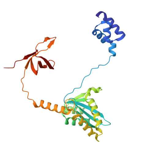
Gag-Pol polyprotein281Visna/maedi virus EV1 KV1772Mutation(s): 0 
Gene Names: pol
UniProt
Find proteins for P35956 (Maedi visna virus (strain KV1772))
Explore P35956 
Go to UniProtKB:  P35956
Entity Groups
Sequence Clusters30% Identity50% Identity70% Identity90% Identity95% Identity100% Identity
UniProt GroupP35956
Sequence Annotations
Expand
Reference Sequence
Find similar proteins by:|  3D Structure
Entity ID: 4
MoleculeChains  Sequence LengthOrganismDetailsImage
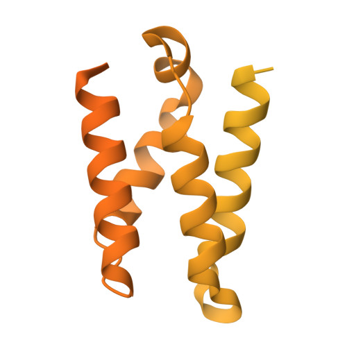
PC4 and SFRS1-interacting protein530Homo sapiensMutation(s): 0 
Gene Names: PSIP1, DFS70, LEDGF, PSIP2
UniProt & NIH Common Fund Data Resources
Find proteins for O75475 (Homo sapiens)
Explore O75475 
Go to UniProtKB:  O75475
PHAROS:  O75475
GTEx:  ENSG00000164985 
Entity Groups
Sequence Clusters30% Identity50% Identity70% Identity90% Identity95% Identity100% Identity
UniProt GroupO75475
Sequence Annotations
Expand
Reference Sequence
Find similar nucleic acids by:  Sequence
Entity ID: 2
MoleculeChains LengthOrganismImage
EV306I [auth W],
Y
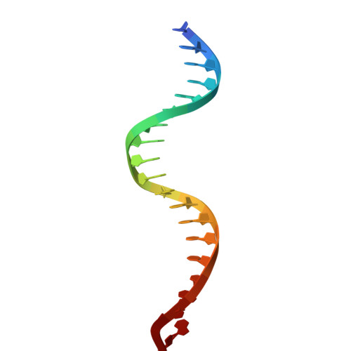
21DNA molecule
Sequence Annotations
Expand
Reference Sequence
Find similar nucleic acids by:  Sequence
Entity ID: 3
MoleculeChains LengthOrganismImage
EV272J [auth X],
Z
19DNA molecule
Sequence Annotations
Expand
Reference Sequence

Small Molecules

Ligands 1 Unique
IDChains Name / Formula / InChI Key2D Diagram3D Interactions
ZN

Query on ZN


Download:Ideal Coordinates CCD File
GA [auth A]
HA [auth B]
IA [auth C]
JA [auth D]
KA [auth E]
GA [auth A],
HA [auth B],
IA [auth C],
JA [auth D],
KA [auth E],
LA [auth F],
MA [auth G],
NA [auth H],
OA [auth I],
PA [auth J],
QA [auth K],
RA [auth L],
SA [auth M],
TA [auth N],
UA [auth O],
VA [auth P]
ZINC ION
Zn
PTFCDOFLOPIGGS-UHFFFAOYSA-N

Experimental Data & Validation

Experimental Data

  • Method: ELECTRON MICROSCOPY
  • Resolution: 2.80 Å
  • Aggregation State: PARTICLE 
  • Reconstruction Method: SINGLE PARTICLE 
EM Software:
TaskSoftware PackageVersion
RECONSTRUCTIONcryoSPARC4.5.3
MODEL REFINEMENTPHENIX1.21.2_5419

Structure Validation

View Full Validation Report


Entry History 

& Funding Information

Deposition Data


Funding OrganizationLocationGrant Number
National Institutes of Health/National Institute Of Allergy and Infectious Diseases (NIH/NIAID)United StatesU54AI170791
Wellcome TrustUnited KingdomCC2058
Medical Research Council (MRC, United Kingdom)United KingdomCC2058
Cancer Research UKUnited KingdomCC2058

Revision History  (Full details and data files)

  • Version 1.0: 2026-07-29
    Type: Initial release
  • Version 1.1: 2026-09-30
    Changes: Data collection, Database references