Integrated structural dynamics uncover a new B 12 photoreceptor activation mode.
Rios-Santacruz, R., Poddar, H., Pounot, K., Heyes, D.J., Coquelle, N., Mackintosh, M.J., Johannissen, L.O., Schianchi, S., Jeffreys, L.N., De Zitter, E., Munro, R., Appleby, M., Axford, D., Beale, E.V., Cliff, M.J., Davila-Miliani, M.C., Engilberge, S., Gotthard, G., Hadjidemetriou, K., Hardman, S.J.O., Horrell, S., Hub, J.S., Ishihara, K., Jaho, S., Karras, G., Kataoka, M., Kawakami, R., Mason, T., Okumura, H., Owada, S., Owen, R.L., Royant, A., Saaret, A., Sakuma, M., Shanmugam, M., Sugimoto, H., Tono, K., Zala, N., Beale, J.H., Tosha, T., Colletier, J.P., Levantino, M., Hay, S., Kozlowski, P.M., Leys, D., Scrutton, N.S., Weik, M., Schiro, G.(2026) Nature 650: 1045-1052
- PubMed: 41639453 Search on PubMedSearch on PubMed Central
- DOI: https://doi.org/10.1038/s41586-025-10074-2
- Primary Citation Related Structures:
9S06, 9S07, 9S08, 9S09, 9S0A, 9S0B, 9S0C, 9S0D, 9S0E, 9S0F, 9S0G, 9S0H, 9S0I, 9S0J - PubMed Abstract:
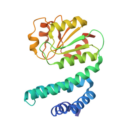
Photoreceptor proteins regulate fundamental biological processes such as vision, photosynthesis and circadian rhythms 1 . A large photoreceptor subfamily uses vitamin B 12 derivatives for light sensing 2 , contrasting with the well-established mode of action of these organometallic derivatives in thermally activated enzymatic reactions 3 . The exact molecular mechanism of B 12 photoreception and how this differs from the thermal pathways remains unknown. Here we provide a detailed description of photoactivation in the prototypical B 12 photoreceptor CarH 4,5 from nanoseconds to seconds, combining time-resolved and temperature-resolved structural and spectroscopic methods with quantum chemical calculations. Building on the crystal structures of the initial tetrameric dark and final monomeric light-activated states 5 , our structural snapshots of key intermediates in the truncated B 12 -binding domain illustrate how photocleavage of a cobalt-carbon (Co-C) bond within the B 12 chromophore adenosylcobalamin triggers a series of structural changes that propagate throughout CarH. Breakage of the photolabile Co-C5' bond leads to the formation of a previously unknown adduct that links the C4' position of the adenosyl moiety to the Co ion and can subsequently be cleaved thermally over longer timescales to allow release of the adenosyl group, ultimately causing tetramer dissociation 4,5 . This adduct, which differentiates CarH from thermally activated B 12 enzymes, steers the photoactivation pathway and acts as the molecular bridge between photochemical and photobiological timescales. The biological relevance of our study is corroborated by kinetic data on full-length CarH in the presence of DNA. Our results offer a spatiotemporal understanding of CarH photoactivation and pave the way for designing B 12 -dependent photoreceptors for optogenetic applications.
- University of Grenoble Alpes, CEA, CNRS, Institut de Biologie Structurale, Grenoble, France.
Organizational Affiliation: