9RW2 | pdb_00009rw2

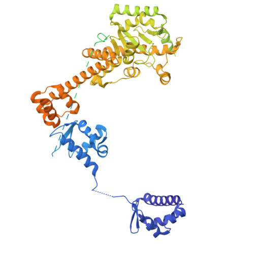
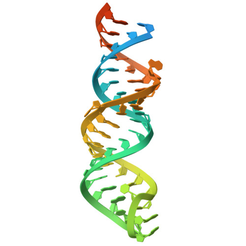
Egalitarian-BicD in complex with K10 TLS RNA (Structure E)


Experimental Data Snapshot

  • Method: ELECTRON MICROSCOPY
  • Resolution: 3.60 Å
  • Aggregation State: PARTICLE 
  • Reconstruction Method: SINGLE PARTICLE 

Starting Model: in silico
View more details

wwPDB Validation 3D Report Full Report

Validation slider image for 9RW2

This is version 1.1 of the entry. See complete history. 

Literature

Structural basis for recognition of diverse localizing mRNAs by Egl-BicD.

Singh, K., Chilaeva, S., McClintock, M.A., Carter, A.P., Bullock, S.L.

(2026) Nat Struct Mol Biol 33: 882-893

  • DOI: https://doi.org/10.1038/s41594-026-01794-8
  • Primary Citation Related Structures: 
    9RVY, 9RVZ, 9RW0, 9RW1, 9RW2, 9RW3, 9RW4, 9RW5, 9RW6, 9RW7, 9RW8, 9RW9, 9RWA, 9RWB

  • PubMed Abstract: 

    Localization of mRNAs is a widespread mechanism for dictating where proteins operate in cells and underpins many fundamental processes, from embryonic patterning to synaptic plasticity. This spatial control is mediated by the interaction of 'localization signals' in target mRNAs with RNA-binding proteins (RBPs). These signals frequently lack overt sequence or structural patterns, raising the question of how specificity is achieved. Here we investigate this issue using the Drosophila RBP Egalitarian (Egl), which couples mRNAs to microtubule-based transport through Bicaudal D (BicD) and the dynein motor. We present cryo-electron microscopy structures of Egl-BicD bound to six different RNAs. Egl uses multiple noncanonical double-stranded RNA-binding domains to cooperatively form a recognition pocket around localization signals. Despite substantial variation in length and sequence, each signal adopts a bent stem-loop conformation that, together with base-pair identities at two defined sites, drives Egl engagement. We further demonstrate that Egl dimers couple RNA binding to transport initiation through coincident detection of two RNA elements within the same transcript. Thus, localizing mRNAs are recognized through a combination of shape, positional sequence features and number of structured RNA elements. This work reveals a molecular strategy by which diverse mRNAs can be selectively engaged by a single RBP.


  • Organizational Affiliation: 
    • Division of Structural Studies, MRC Laboratory of Molecular Biology, Cambridge, UK.

Macromolecule Content 

  • Total Structure Weight: 442.47 kDa 
  • Atom Count: 8,127 
  • Modeled Residue Count: 1,116 
  • Deposited Residue Count: 3,708 
  • Unique protein chains: 2
  • Unique nucleic acid chains: 1

Macromolecules


Find similar proteins by:|  3D Structure
Entity ID: 1
MoleculeChains  Sequence LengthOrganismDetailsImage
Protein bicaudal D
A, B
782Drosophila melanogasterMutation(s): 0 
Gene Names: BicD, CG6605
UniProt
Find proteins for P16568 (Drosophila melanogaster)
Explore P16568 
Go to UniProtKB:  P16568
Entity Groups
Sequence Clusters30% Identity50% Identity70% Identity90% Identity95% Identity100% Identity
UniProt GroupP16568
Sequence Annotations
Expand
Reference Sequence
Find similar proteins by:|  3D Structure
Entity ID: 2
MoleculeChains  Sequence LengthOrganismDetailsImage
Egalitarian, isoform BC [auth D],
D [auth C]
1,012Drosophila melanogasterMutation(s): 0 
Gene Names: egl, Dmel\CG4051, egal, EGL, CG4051, Dmel_CG4051
UniProt
Find proteins for Q9W1K4 (Drosophila melanogaster)
Explore Q9W1K4 
Go to UniProtKB:  Q9W1K4
Entity Groups
Sequence Clusters30% Identity50% Identity70% Identity90% Identity95% Identity100% Identity
UniProt GroupQ9W1K4
Sequence Annotations
Expand
Reference Sequence
Find similar nucleic acids by:  (by identity cutoff) 
Entity ID: 3
MoleculeChains LengthOrganismImage
TLS
E, F
60Drosophila melanogaster
Sequence Annotations
Expand
Reference Sequence

Experimental Data & Validation

Experimental Data

  • Method: ELECTRON MICROSCOPY
  • Resolution: 3.60 Å
  • Aggregation State: PARTICLE 
  • Reconstruction Method: SINGLE PARTICLE 
EM Software:
TaskSoftware PackageVersion
MODEL REFINEMENTPHENIX1.20_4459
RECONSTRUCTIONRELION5

Structure Validation

View Full Validation Report


Entry History 

& Funding Information

Deposition Data


Funding OrganizationLocationGrant Number
Medical Research Council (MRC, United Kingdom)United KingdomMC_UP_A025_1011
European Molecular Biology Organization (EMBO)European UnionALTF 197-2021

Revision History  (Full details and data files)

  • Version 1.0: 2026-03-25
    Type: Initial release
  • Version 1.1: 2026-10-14
    Changes: Data collection, Database references