9RPR | pdb_00009rpr

Cryo-EM structure of LptDEM complex containing Shigella flexneri LptE and endogenous E. coli LptD and LptM


Experimental Data Snapshot

  • Method: ELECTRON MICROSCOPY
  • Resolution: 2.78 Å
  • Aggregation State: PARTICLE 
  • Reconstruction Method: SINGLE PARTICLE 

Starting Model: in silico
View more details

wwPDB Validation   3D Report Full Report


This is version 1.0 of the entry. See complete history


Literature

Small siphophage binding to an open state of the LptDE outer membrane lipopolysaccharide translocon.

Dunbar, E.Clark, R.Basle, A.Allyjaun, S.Newman, H.Hubbard, J.Khalid, S.van den Berg, B.

(2025) Proc Natl Acad Sci U S A 122: e2516650122-e2516650122

  • DOI: https://doi.org/10.1073/pnas.2516650122
  • Primary Citation of Related Structures:  
    9RPR, 9RPS, 9RPT, 9RPW, 9RQI

  • PubMed Abstract: 

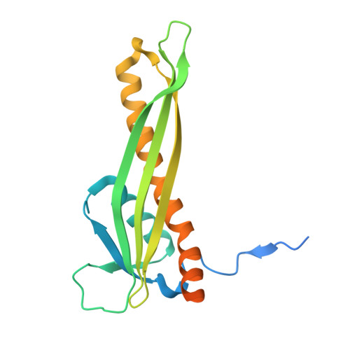
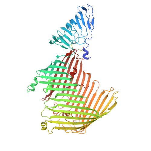
    Bacteriophages are bacterial viruses that provide alternatives to small-molecule drugs to combat infections by antibiotic-resistant bacteria. To infect a bacterial host, a phage needs to bind to the bacterial surface via receptor binding proteins (RBPs), which are critical for determining host specificity. For functionally important receptors, the RBP-receptor interaction could be exploited via phage steering, where emerging bacterial resistance due to receptor modification could make bacteria less fit or virulent. Despite this, relatively little is known about RBP-receptor interactions. Here, we build on the recent discovery of coliphages that have the outer membrane (OM) lipopolysaccharide translocon LptDE as their terminal receptor and show via cryogenic electron microscopy that, surprisingly, the RBP of the small siphophage Oekolampad binds to a hitherto unobserved, open state of LptDE. The open lateral gate of LptD is occupied by a β-strand peptide originating from the degraded N-terminal jellyroll domain of LptD, suggesting the possibility of LptD inhibition via peptidomimetics. A structure of LptDE in complex with the superinfection exclusion (SE) protein Rtp45 of the Oekolampad-related phage Rtp shows a mechanism of SE where Rtp45-induced conformational changes in LptD resulting from steric clashes preclude RBP binding. Finally, analysis of spontaneous Oekolampad-resistant Escherichia coli mutants identifies mutations in LptD that abolish the LptDE-RBP interaction in vitro. SDS-EDTA sensitivity assays of the mutants show no major OM defects, consistent with largely preserved LptDE function, and suggesting that phage steering via LptDE might be challenging.


  • Organizational Affiliation
    • Biosciences Institute, The Medical School, Newcastle University, Newcastle upon Tyne NE2 4HH, United Kingdom.

Macromolecules
Find similar proteins by:  (by identity cutoff)  |  3D Structure
Entity ID: 1
MoleculeChains Sequence LengthOrganismDetailsImage
LPS-assembly lipoprotein LptEA [auth B]199Shigella flexneriMutation(s): 0 
Gene Names: lptErlpBSF0640S0662
UniProt
Find proteins for Q83LX4 (Shigella flexneri)
Explore Q83LX4 
Go to UniProtKB:  Q83LX4
Entity Groups  
Sequence Clusters30% Identity50% Identity70% Identity90% Identity95% Identity100% Identity
UniProt GroupQ83LX4
Sequence Annotations
Expand
  • Reference Sequence
Find similar proteins by:  (by identity cutoff)  |  3D Structure
Entity ID: 2
MoleculeChains Sequence LengthOrganismDetailsImage
LPS-assembly protein LptDB [auth A]784Escherichia coliMutation(s): 0 
UniProt
Find proteins for P31554 (Escherichia coli (strain K12))
Explore P31554 
Go to UniProtKB:  P31554
Entity Groups  
Sequence Clusters30% Identity50% Identity70% Identity90% Identity95% Identity100% Identity
UniProt GroupP31554
Sequence Annotations
Expand
  • Reference Sequence
Find similar proteins by:  (by identity cutoff)  |  3D Structure
Entity ID: 3
MoleculeChains Sequence LengthOrganismDetailsImage
LPS-assembly lipoprotein LptM48Escherichia coliMutation(s): 0 
UniProt
Find proteins for P0ADN6 (Escherichia coli (strain K12))
Explore P0ADN6 
Go to UniProtKB:  P0ADN6
Entity Groups  
Sequence Clusters30% Identity50% Identity70% Identity90% Identity95% Identity100% Identity
UniProt GroupP0ADN6
Sequence Annotations
Expand
  • Reference Sequence
Experimental Data & Validation

Experimental Data

  • Method: ELECTRON MICROSCOPY
  • Resolution: 2.78 Å
  • Aggregation State: PARTICLE 
  • Reconstruction Method: SINGLE PARTICLE 
EM Software:
TaskSoftware PackageVersion
MODEL REFINEMENTCoot

Structure Validation

View Full Validation Report


Entry History & Funding Information

Deposition Data


Funding OrganizationLocationGrant Number
Medical Research Council (MRC, United Kingdom)United KingdomU-016275 IAA-CiC
Other private--

Revision History  (Full details and data files)

  • Version 1.0: 2025-12-10
    Type: Initial release