9RMX | pdb_00009rmx

CryoEM reconstruction of integrase filament at the lumen of native HIV-1 cores (box size 34.2 nm)


Experimental Data Snapshot

  • Method: ELECTRON MICROSCOPY
  • Resolution: 4.63 Å
  • Aggregation State: PARTICLE 
  • Reconstruction Method: SINGLE PARTICLE 

Starting Models: experimental
View more details

wwPDB Validation 3D Report Full Report

Validation slider image for 9RMX

This is version 1.2 of the entry. See complete history

Literature

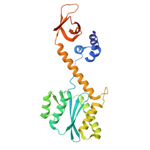
Integrase anchors viral RNA to the HIV-1 capsid interior.

Singer, M.R.Li, Z.Rey, J.S.Hope, J.Chenavier, F.Cook, N.J.Punch, E.Smith, J.Zhou, Z.Maslen, S.Masino, L.Nans, A.Skehel, M.Taylor, I.A.Zanetti, G.Zhang, P.Perilla, J.R.Engelman, A.N.Cherepanov, P.

(2026) Nature 

  • DOI: https://doi.org/10.1038/s41586-026-10154-x
  • Primary Citation Related Structures: 
    9RMU, 9RMX

  • PubMed Abstract: 

    HIV-1 integrase (IN) promotes encapsulation of viral genomic RNA into mature viral cores, and this function is a target for ongoing antiretroviral drug development efforts 1-3 . Here we determined the cryogenic electron microscopy (cryo-EM) structure of a primate lentiviral IN in a complex with RNA, revealing a linear filament made of IN octamer repeat units, each comprising a pair of asymmetric homotetramers. The assembly is stabilized through IN-RNA interactions involving mainly the IN C-terminal domains and RNA backbone. The spacing and orientation of the IN filament repeat units closely matched those of consecutive capsid (CA) hexamers within the mature CA lattice. Using cryo-EM images of native purified HIV-1 cores, we refined the structure of the IN filament as it propagates along the luminal side of the CA lattice. Each IN tetramer within the filament nestled in a CA hexamer, engaging closely with the major homology regions. Substitutions of residues involved in IN-CA contacts yielded eccentric virions with RNA nucleoids located outside of the cores. Collectively, our results establish the structural basis for the HIV-1 IN-RNA interaction and reveal that IN forms an RNA-binding module on the luminal side of the mature CA lattice.


  • Organizational Affiliation
    • Chromatin Structure & Mobile DNA Laboratory, The Francis Crick Institute, London, UK.

Macromolecule Content 

  • Total Structure Weight: 1,213.15 kDa 
  • Atom Count: 72,751 
  • Modeled Residue Count: 9,257 
  • Deposited Residue Count: 10,848 
  • Unique protein chains: 2

Macromolecules

Find similar proteins by:|  3D Structure
Entity ID: 1
MoleculeChains  Sequence LengthOrganismDetailsImage
Integrase
A, B, C, D, E
A, B, C, D, E, F, G, H, I, J, K, L
288Human immunodeficiency virus 1Mutation(s): 2 
Gene Names: gag-pol
EC: 2.7.7 (PDB Primary Data), 3.1 (PDB Primary Data)
UniProt
Find proteins for P12497 (Human immunodeficiency virus type 1 group M subtype B (isolate NY5))
Explore P12497 
Go to UniProtKB:  P12497
Entity Groups
Sequence Clusters30% Identity50% Identity70% Identity90% Identity95% Identity100% Identity
UniProt GroupP12497
Sequence Annotations
Expand
Reference Sequence
Find similar proteins by:|  3D Structure
Entity ID: 2
MoleculeChains  Sequence LengthOrganismDetailsImage
Gag polyprotein231Human immunodeficiency virus 1Mutation(s): 0 
Gene Names: gag
UniProt
Find proteins for Q72497 (Human immunodeficiency virus type 1)
Explore Q72497 
Go to UniProtKB:  Q72497
Entity Groups
Sequence Clusters30% Identity50% Identity70% Identity90% Identity95% Identity100% Identity
UniProt GroupQ72497
Sequence Annotations
Expand
Reference Sequence

Small Molecules

Ligands 2 Unique
IDChains Name / Formula / InChI Key2D Diagram3D Interactions
IHP

Query on IHP


Download:Ideal Coordinates CCD File
EB [auth X]
FB [auth Z]
GB [auth a]
HB [auth b]
IB [auth g]
EB [auth X],
FB [auth Z],
GB [auth a],
HB [auth b],
IB [auth g],
JB [auth i],
KB [auth m],
LB [auth q]
INOSITOL HEXAKISPHOSPHATE
C6 H18 O24 P6
IMQLKJBTEOYOSI-GPIVLXJGSA-N
ZN

Query on ZN


Download:Ideal Coordinates CCD File
AB [auth I]
BB [auth J]
CB [auth K]
DB [auth L]
SA [auth A]
AB [auth I],
BB [auth J],
CB [auth K],
DB [auth L],
SA [auth A],
TA [auth B],
UA [auth C],
VA [auth D],
WA [auth E],
XA [auth F],
YA [auth G],
ZA [auth H]
ZINC ION
Zn
PTFCDOFLOPIGGS-UHFFFAOYSA-N

Experimental Data & Validation

Experimental Data

  • Method: ELECTRON MICROSCOPY
  • Resolution: 4.63 Å
  • Aggregation State: PARTICLE 
  • Reconstruction Method: SINGLE PARTICLE 
EM Software:
TaskSoftware PackageVersion
RECONSTRUCTIONcryoSPARC4.6.2
MODEL REFINEMENTPHENIX1.21.2_5419

Structure Validation

View Full Validation Report


Entry History 

& Funding Information

Deposition Data


Funding OrganizationLocationGrant Number
National Institutes of Health/National Institute Of Allergy and Infectious Diseases (NIH/NIAID)United StatesU54AI170791
Wellcome TrustUnited KingdomCC2058
Medical Research Council (MRC, United Kingdom)United KingdomCC2058
Cancer Research UKUnited KingdomCC2058

Revision History  (Full details and data files)

  • Version 1.0: 2025-12-10
    Type: Initial release
  • Version 1.1: 2026-02-18
    Changes: Data collection, Database references
  • Version 1.2: 2026-03-04
    Changes: Data collection, Database references