9RD5 | pdb_00009rd5

Phi3T SroF bound to DNA


Experimental Data Snapshot

  • Method: X-RAY DIFFRACTION
  • Resolution: 2.50 Å
  • R-Value Free: 
    0.253 (Depositor), 0.253 (DCC) 
  • R-Value Work: 
    0.207 (Depositor), 0.207 (DCC) 
  • R-Value Observed: 
    0.209 (Depositor) 

Starting Model: experimental
View more details

wwPDB Validation 3D Report Full Report

Validation slider image for 9RD5

This is version 1.1 of the entry. See complete history

Literature

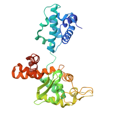
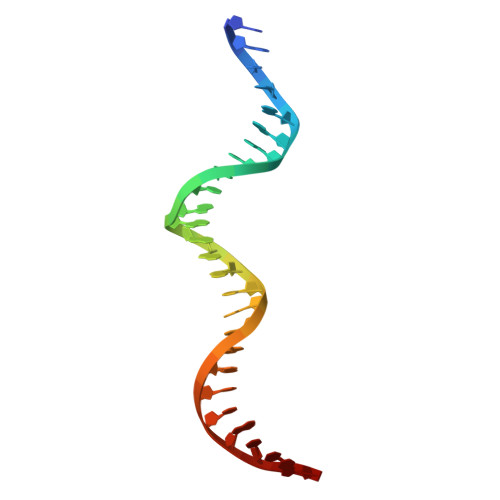
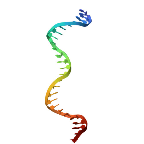
A DNA recognition-mimicry switch governs induction in arbitrium phages.

Chmielowska, C.Zamora-Caballero, S.Mancheno-Bonillo, J.Li, Y.Sin, D.Borenstein, T.Bendori, S.O.Eldar, A.Marina, A.Penades, J.R.

(2026) Cell Host Microbe 34: 291-303.e10

  • DOI: https://doi.org/10.1016/j.chom.2026.01.012
  • Primary Citation Related Structures: 
    9RD5, 9RGL

  • PubMed Abstract: 

    Temperate phages integrate multiple information sources to regulate lysis-lysogeny transitions. SPbeta-like phages use arbitrium signaling and DNA damage to control repressor activity during lytic induction, but how the repressor functions and is inactivated by the SOS response remains unclear. Here, we show that SroF, the SPbeta-like phage repressor, binds DNA via a mechanism involving its integrase-like fold, enabling stable prophage repression. Upon DNA damage, the host SOS response triggers derepression of an antirepressor, Sar. Sar binds SroF by mimicking the DNA structure recognized by the repressor, thereby inactivating its function and inducing phage. This mechanism is conserved across SPbeta-like phages, which encode multiple, specific SroF-Sar pairs. Surprisingly, repressor inactivation alone is insufficient for efficient induction when arbitrium levels are high. Our results uncover the mechanism underlying a double layer of control that ensures phage induction occurs only under SOS conditions and in the absence of neighboring prophages.


  • Organizational Affiliation
    • Department of Infectious Disease, Imperial College London, London SW7 2AZ, UK; Centre for Bacterial Resistance Biology, Imperial College London, London SW7 2AZ, UK.

Macromolecule Content 

  • Total Structure Weight: 54.42 kDa 
  • Atom Count: 3,809 
  • Modeled Residue Count: 369 
  • Deposited Residue Count: 377 
  • Unique protein chains: 1
  • Unique nucleic acid chains: 2

Macromolecules


Find similar proteins by:|  3D Structure
Entity ID: 3
MoleculeChains  Sequence LengthOrganismDetailsImage
Core-binding (CB) domain-containing proteinC [auth H]325Bacillus phage phi3TMutation(s): 0 
Gene Names: phi3T_97
Find similar nucleic acids by:  (by identity cutoff) 
Entity ID: 1
MoleculeChains LengthOrganismImage
DNA (26-MER)26Bacillus phage phi3T
Sequence Annotations
Expand
Reference Sequence
Find similar nucleic acids by:  (by identity cutoff) 
Entity ID: 2
MoleculeChains LengthOrganismImage
DNA (26-MER)26Bacillus phage phi3T
Sequence Annotations
Expand
Reference Sequence

Small Molecules

Experimental Data & Validation

Experimental Data

  • Method: X-RAY DIFFRACTION
  • Resolution: 2.50 Å
  • R-Value Free:  0.253 (Depositor), 0.253 (DCC) 
  • R-Value Work:  0.207 (Depositor), 0.207 (DCC) 
  • R-Value Observed: 0.209 (Depositor) 
Space Group: C 2 2 21
Unit Cell:
Length ( Å )Angle ( ˚ )
a = 75.182α = 90
b = 115.618β = 90
c = 154.372γ = 90
Software Package:
Software NamePurpose
MxCuBEdata collection
PHENIXrefinement
MOSFLMdata reduction
Aimlessdata scaling
PHASERphasing

Structure Validation

View Full Validation Report


Entry History 

& Funding Information

Deposition Data


Funding OrganizationLocationGrant Number
European Research Council (ERC)European Union101118890
Ministerio de Ciencia e Innovacion (MCIN)SpainPID2022-137201NB-I00
Generalitat ValencianaSpainCIPROM/2023/30

Revision History  (Full details and data files)

  • Version 1.0: 2026-01-14
    Type: Initial release
  • Version 1.1: 2026-04-22
    Changes: Database references, Structure summary