9RC2 | pdb_00009rc2

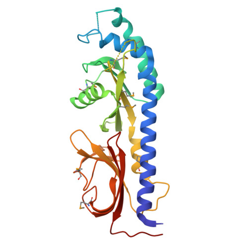
DvhD, periplasmic domain


Experimental Data Snapshot

  • Method: X-RAY DIFFRACTION
  • Resolution: 3.00 Å
  • R-Value Free: 
    0.302 (Depositor), 0.319 (DCC) 
  • R-Value Work: 
    0.270 (Depositor), 0.289 (DCC) 
  • R-Value Observed: 
    0.273 (Depositor) 

wwPDB Validation 3D Report Full Report

Validation slider image for 9RC2

This is version 1.1 of the entry. See complete history

Literature

Structural analyses uncover protease-adhesin interactions and c-di-GMP receptor regulation in sulfate-reducing bacteria.

Font, M.E.Karbelkar, A.A.Lormand, J.D.Mortensen, S.Garcia-Garcia, M.J.O'Toole, G.A.Sondermann, H.

(2026) Nat Commun 17

  • DOI: https://doi.org/10.1038/s41467-026-71936-5
  • Primary Citation Related Structures: 
    9RBZ, 9RC0, 9RC2

  • PubMed Abstract: 

    Desulfovibrio vulgaris is a sulfate-reducing organism with biofim-forming capacity relevant for bioremediation and microbe-induced corrosion. Biofilm formation of D. vulgaris depends on two large adhesins that are regulated by proteins encoded in the Dvh operon, which resembles the gammaproteobacterial Lap system in composition but differs in the sequence and domain organization of its regulatory proteins, DvhG and DvhD. We show that DvhG is a calcium-dependent protease that targets the periplasmic domains of both adhesins via extensive interactions. Additionally, structures of DvhD establish this HD-GYP domain-containing protein as a c-di-GMP-dependent switch with a periplasmic dCache domain. Our data support a model in which DvhD controls DvhG activity through a c-di-GMP-dependent mechanism that is molecularly distinct, but functionally analogous to LapD. Together, our results reveal how conserved regulatory logic can be implemented through distinct molecular architectures, highlighting the evolutionary flexibility of c-di-GMP signaling networks in controlling surface attachment across diverse bacterial lineages.


  • Organizational Affiliation
    • CSSB Centre for Structural Systems Biology, Deutsches Elektronen-Synchrotron DESY, Hamburg, Germany.

Macromolecule Content 

  • Total Structure Weight: 29.6 kDa 
  • Atom Count: 1,881 
  • Modeled Residue Count: 252 
  • Deposited Residue Count: 271 
  • Unique protein chains: 1

Macromolecules

Find similar proteins by:|  3D Structure
Entity ID: 1
MoleculeChains  Sequence LengthOrganismDetailsImage
HD domain/sensory box protein271Nitratidesulfovibrio vulgaris str. HildenboroughMutation(s): 0 
Gene Names: DVU_1020
UniProt
Find proteins for Q72DA9 (Nitratidesulfovibrio vulgaris (strain ATCC 29579 / DSM 644 / CCUG 34227 / NCIMB 8303 / VKM B-1760 / Hildenborough))
Explore Q72DA9 
Go to UniProtKB:  Q72DA9
Entity Groups
Sequence Clusters30% Identity50% Identity70% Identity90% Identity95% Identity100% Identity
UniProt GroupQ72DA9
Sequence Annotations
Expand
Reference Sequence

Small Molecules

Modified Residues  1 Unique
IDChains TypeFormula2D DiagramParent
MSE
Query on MSE
A
L-PEPTIDE LINKINGC5 H11 N O2 SeMET

Experimental Data & Validation

Experimental Data

  • Method: X-RAY DIFFRACTION
  • Resolution: 3.00 Å
  • R-Value Free:  0.302 (Depositor), 0.319 (DCC) 
  • R-Value Work:  0.270 (Depositor), 0.289 (DCC) 
  • R-Value Observed: 0.273 (Depositor) 
Space Group: P 64 2 2
Unit Cell:
Length ( Å )Angle ( ˚ )
a = 120.38α = 90
b = 120.38β = 90
c = 123.958γ = 120
Software Package:
Software NamePurpose
PHENIXrefinement
XDSdata reduction
SCALAdata scaling
PHENIXphasing

Structure Validation

View Full Validation Report


Entry History 

& Funding Information

Deposition Data


Funding OrganizationLocationGrant Number
National Institutes of Health/National Institute Of Allergy and Infectious Diseases (NIH/NIAID)United States1R01AI168017

Revision History  (Full details and data files)

  • Version 1.0: 2026-03-04
    Type: Initial release
  • Version 1.1: 2026-04-29
    Changes: Database references