9RBR | pdb_00009rbr

Semliki Forest virus trimer 2 in complex with ApoER2 LA5


Experimental Data Snapshot

  • Method: ELECTRON MICROSCOPY
  • Resolution: 2.70 Å
  • Aggregation State: PARTICLE 
  • Reconstruction Method: SINGLE PARTICLE 

wwPDB Validation   3D Report Full Report


This is version 1.0 of the entry. See complete history


Literature

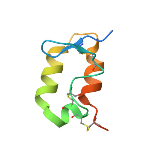
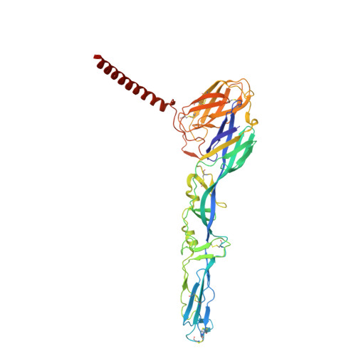
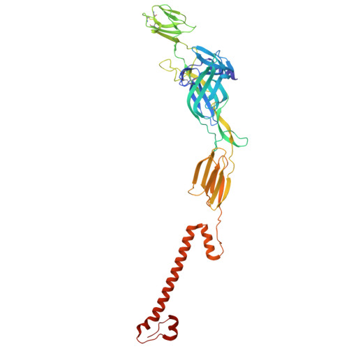
Molecular basis of ApoER2-mediated Semliki Forest virus entry.

Du, B.Song, X.Zhao, B.Shi, Z.Liu, Z.Wang, S.Wei, L.He, X.Huiskonen, J.T.Yang, D.Wang, J.

(2025) Nat Commun 

  • DOI: https://doi.org/10.1038/s41467-025-67550-6
  • Primary Citation of Related Structures:  
    9RBQ, 9RBR

  • PubMed Abstract: 

    The very low-density lipoprotein receptor (VLDLR) and apolipoprotein E receptor 2 (ApoER2) serve as entry receptors for the Semliki Forest virus (SFV). VLDLR interacts with the SFV E1 domain III (DIII) through multiple LDLR class A (LA) domains. However, the ApoER2-mediated SFV entry mechanism remains unclear. Here, we perform biochemical and cellular results and determine the cryogenic electron microscopy (cryo-EM) structures of SFV complexed with ApoER2 LA5 and full-length ApoER2, demonstrating that among the seven LA domains of ApoER2 isoform 1, only LA5 specifically binds to the SFV E1-DIII via a limited interface (353 Ų) and facilitates cell attachment and entry. Site-directed mutagenesis confirms the significance of the residues at the SFV-ApoER2 interface. Significantly, a soluble LA5 decoy receptor neutralizes SFV infection and protects mice from lethal SFV challenge. These findings reveal a LA5-dependent receptor engagement mechanism for SFV entry via ApoER2, distinct from VLDLR.


  • Organizational Affiliation
    • State Key Laboratory for Animal Disease Control and Prevention & Data Center for Field Scientific Observation and Research of Animal Diseases, Ministry of Agriculture and Rural Affair, Harbin Veterinary Research Institute, Chinese Academy of Agricultural Sciences, Harbin, PR China.

Macromolecules
Find similar proteins by:  (by identity cutoff)  |  3D Structure
Entity ID: 1
MoleculeChains Sequence LengthOrganismDetailsImage
Protein E3A [auth D],
B [auth E],
C [auth F]
66Semliki Forest virusMutation(s): 0 
UniProt
Find proteins for P0DJZ6 (Semliki forest virus)
Explore P0DJZ6 
Go to UniProtKB:  P0DJZ6
Entity Groups  
Sequence Clusters30% Identity50% Identity70% Identity90% Identity95% Identity100% Identity
UniProt GroupP0DJZ6
Sequence Annotations
Expand
  • Reference Sequence
Find similar proteins by:  (by identity cutoff)  |  3D Structure
Entity ID: 2
MoleculeChains Sequence LengthOrganismDetailsImage
Structural polyproteinD [auth G],
E [auth H],
F [auth I]
438Semliki Forest virusMutation(s): 0 
UniProt
Find proteins for A0A0F6PP03 (Semliki forest virus)
Explore A0A0F6PP03 
Go to UniProtKB:  A0A0F6PP03
Entity Groups  
Sequence Clusters30% Identity50% Identity70% Identity90% Identity95% Identity100% Identity
UniProt GroupA0A0F6PP03
Glycosylation
Glycosylation Sites: 1
Sequence Annotations
Expand
  • Reference Sequence
Find similar proteins by:  (by identity cutoff)  |  3D Structure
Entity ID: 3
MoleculeChains Sequence LengthOrganismDetailsImage
Envelope glycoprotein E2G [auth J],
H [auth K],
I [auth L]
422Semliki Forest virusMutation(s): 0 
UniProt
Find proteins for P0DJZ6 (Semliki forest virus)
Explore P0DJZ6 
Go to UniProtKB:  P0DJZ6
Entity Groups  
Sequence Clusters30% Identity50% Identity70% Identity90% Identity95% Identity100% Identity
UniProt GroupP0DJZ6
Glycosylation
Glycosylation Sites: 2
Sequence Annotations
Expand
  • Reference Sequence
Find similar proteins by:  (by identity cutoff)  |  3D Structure
Entity ID: 4
MoleculeChains Sequence LengthOrganismDetailsImage
Capsid proteinJ [auth M],
K [auth N],
L [auth O]
267Semliki Forest virusMutation(s): 0 
EC: 3.4.21.90
UniProt
Find proteins for P0DJZ6 (Semliki forest virus)
Explore P0DJZ6 
Go to UniProtKB:  P0DJZ6
Entity Groups  
Sequence Clusters30% Identity50% Identity70% Identity90% Identity95% Identity100% Identity
UniProt GroupP0DJZ6
Sequence Annotations
Expand
  • Reference Sequence
Oligosaccharides

Help

Entity ID: 5
MoleculeChains Length2D Diagram Glycosylation3D Interactions
beta-D-mannopyranose-(1-4)-2-acetamido-2-deoxy-beta-D-glucopyranose-(1-4)-[alpha-D-mannopyranose-(1-6)]2-acetamido-2-deoxy-beta-D-glucopyranoseM [auth A],
N [auth B],
O [auth C]
4N-Glycosylation
Entity ID: 6
MoleculeChains Length2D Diagram Glycosylation3D Interactions
2-acetamido-2-deoxy-beta-D-glucopyranose-(1-4)-2-acetamido-2-deoxy-beta-D-glucopyranose-(1-4)-2-acetamido-2-deoxy-beta-D-glucopyranoseP,
R [auth Q],
T [auth R]
3N-Glycosylation
Entity ID: 7
MoleculeChains Length2D Diagram Glycosylation3D Interactions
2-acetamido-2-deoxy-beta-D-glucopyranose-(1-6)-2-acetamido-2-deoxy-beta-D-glucopyranose-(1-4)-2-acetamido-2-deoxy-beta-D-glucopyranose-(1-4)-2-acetamido-2-deoxy-beta-D-glucopyranoseQ [auth d],
S [auth j],
U [auth p]
4N-Glycosylation
Experimental Data & Validation

Experimental Data

  • Method: ELECTRON MICROSCOPY
  • Resolution: 2.70 Å
  • Aggregation State: PARTICLE 
  • Reconstruction Method: SINGLE PARTICLE 
EM Software:
TaskSoftware PackageVersion
RECONSTRUCTIONRELION

Structure Validation

View Full Validation Report


Entry History & Funding Information

Deposition Data


Funding OrganizationLocationGrant Number
Jane and Aatos Erkko FoundationFinland--

Revision History  (Full details and data files)

  • Version 1.0: 2026-01-14
    Type: Initial release