9R8S | pdb_00009r8s

A viral SAVED protein with ring nuclease activity subverts type III CRISPR defence


Experimental Data Snapshot

  • Method: X-RAY DIFFRACTION
  • Resolution: 1.79 Å
  • R-Value Free: 
    0.207 (Depositor), 0.217 (DCC) 
  • R-Value Work: 
    0.165 (Depositor), 0.178 (DCC) 

Starting Model: in silico
View more details

wwPDB Validation 3D Report Full Report

Validation slider image for 9R8S

This is version 1.2 of the entry. See complete history

Literature

A viral SAVED protein with ring nuclease activity degrades the CRISPR second messenger cA4.

Orzechowski, M.Hoikkala, V.Chi, H.McMahon, S.Gloster, T.White, M.F.

(2025) Biochem J 482: 1707-1719

  • DOI: https://doi.org/10.1042/BCJ20253271
  • Primary Citation Related Structures: 
    9R8S

  • PubMed Abstract: 

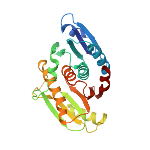
    Type III CRISPR systems typically generate cyclic oligoadenylate second messengers such as cyclic tetra-adenylate (cA4) on detection of foreign RNA. These activate ancillary effector proteins which elicit a diverse range of immune responses. The Calp (CRISPR associated Lon protease) system elicits a transcriptional response to infection when CalpL (Calp Lon protease) binds cA4 in its SAVED (SMODS associated and fused to various effectors domain) sensor domain, resulting in filament formation and activation of the Lon protease domain, which cleaves the anti-Sigma factor CalpT, releasing the CalpS (Calp Sigma factor) for transcriptional remodelling. Here, we show that thermophilic viruses have appropriated the SAVED domain of CalpL as an anti-CRISPR, AcrIII-2 (second anti-CRISPR of type III systems), which they use to degrade cA4. AcrIII-2 dimers sandwich cA4, degrading it in a shared active site to short linear products, using a mechanism highly reminiscent of CalpL. This results in inhibition of a range of cA4 activated effectors in vitro. This is the first example of a virally encoded SAVED domain with ring nuclease activity, highlighting the complex interplay between viruses and cellular defences.


  • Organizational Affiliation
    • School of Biology, University of St Andrews, North Haugh, St Andrews, KY16 9ST, U.K.

Macromolecule Content 

  • Total Structure Weight: 55.45 kDa 
  • Atom Count: 4,025 
  • Modeled Residue Count: 473 
  • Deposited Residue Count: 486 
  • Unique protein chains: 1

Macromolecules

Find similar proteins by:|  3D Structure
Entity ID: 1
MoleculeChains  Sequence LengthOrganismDetailsImage
SMODS-associated and fused to various effectors domain-containing protein
A, B
243Thermocrinis Great Boiling Spring virusMutation(s): 0 

Experimental Data & Validation

Experimental Data

  • Method: X-RAY DIFFRACTION
  • Resolution: 1.79 Å
  • R-Value Free:  0.207 (Depositor), 0.217 (DCC) 
  • R-Value Work:  0.165 (Depositor), 0.178 (DCC) 
Space Group: P 31 2 1
Unit Cell:
Length ( Å )Angle ( ˚ )
a = 110.614α = 90
b = 110.614β = 90
c = 113.027γ = 120
Software Package:
Software NamePurpose
REFMACrefinement
xia2data reduction
xia2data scaling
PHASERphasing

Structure Validation

View Full Validation Report


Entry History 

& Funding Information

Deposition Data


Funding OrganizationLocationGrant Number
European Research Council (ERC)European Union101018608

Revision History  (Full details and data files)

  • Version 1.0: 2025-07-02
    Type: Initial release
  • Version 1.1: 2025-11-26
    Changes: Database references
  • Version 1.2: 2026-01-14
    Changes: Database references