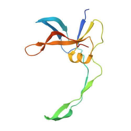
9R7B | pdb_00009r7b

Structure and mechanism of the broad spectrum CRISPR-associated ring nuclease Crn4


Experimental Data Snapshot

  • Method: X-RAY DIFFRACTION
  • Resolution: 1.10 Å
  • R-Value Free: 
    0.185 (Depositor), 0.188 (DCC) 
  • R-Value Work: 
    0.168 (Depositor), 0.177 (DCC) 

Starting Model: in silico
View more details

wwPDB Validation 3D Report Full Report

Validation slider image for 9R7B

This is version 1.2 of the entry. See complete history

Literature

Structure and mechanism of the broad spectrum CRISPR-associated ring nuclease Crn4.

Chi, H.Hoikkala, V.McMahon, S.Graham, S.Gloster, T.White, M.F.

(2025) Nat Commun 17: 889-889

  • DOI: https://doi.org/10.1038/s41467-025-67607-6
  • Primary Citation Related Structures: 
    9QS9, 9R7B, 9SMA

  • PubMed Abstract: 

    Type III CRISPR systems detect the presence of RNA from mobile genetic elements (MGE) in prokaryotes, providing antiviral immunity. On activation, the catalytic Cas10 subunit conjugates ATP to form cyclic oligoadenylate (cOA) signalling molecules that activate ancillary effectors, providing an immune response. Cellular ring nucleases degrade cOA to reset the system. Here, we describe the structure and mechanism of a new family of ring nucleases, Crn4, associated with type III-D CRISPR systems. The crystal structure of Crn4 reveals a small homodimeric protein with a fold unrelated to any known ring nuclease or, indeed, any known protein structure. Crn4 degrades a wide range of cOA species to linear oligoadenylates in vitro and ameliorates type III CRISPR immunity in vivo. Phage and plasmids also encode Crn4 orthologues that may function as anti-CRISPRs. These observations expand our understanding of ring nucleases and reveal a new protein fold for cyclic nucleotide recognition.


  • Organizational Affiliation
    • School of Biology, University of St Andrews, BMS building, North Haugh, St Andrews, Fife, UK.

Macromolecule Content 

  • Total Structure Weight: 13.56 kDa 
  • Atom Count: 993 
  • Modeled Residue Count: 114 
  • Deposited Residue Count: 128 
  • Unique protein chains: 1

Macromolecules

Find similar proteins by:|  3D Structure
Entity ID: 1
MoleculeChains  Sequence LengthOrganismDetailsImage
Crn4b128Hyphomicrobiales bacteriumMutation(s): 0 

Experimental Data & Validation

Experimental Data

  • Method: X-RAY DIFFRACTION
  • Resolution: 1.10 Å
  • R-Value Free:  0.185 (Depositor), 0.188 (DCC) 
  • R-Value Work:  0.168 (Depositor), 0.177 (DCC) 
Space Group: C 1 2 1
Unit Cell:
Length ( Å )Angle ( ˚ )
a = 46.34α = 90
b = 61.33β = 101.95
c = 42.67γ = 90
Software Package:
Software NamePurpose
REFMACrefinement
xia2data reduction
xia2data scaling
PHASERphasing

Structure Validation

View Full Validation Report


Entry History 

& Funding Information

Deposition Data


Funding OrganizationLocationGrant Number
European Research Council (ERC)European Union101018608

Revision History  (Full details and data files)

  • Version 1.0: 2025-12-03
    Type: Initial release
  • Version 1.1: 2026-01-14
    Changes: Database references
  • Version 1.2: 2026-02-04
    Changes: Database references