9R4A | pdb_00009r4a

Spitrobot-2 advances time-resolvedcryo-trapping crystallography to under 25 ms: Human insulin, pH 4.5 (50 ms soaking)


Experimental Data Snapshot

  • Method: X-RAY DIFFRACTION
  • Resolution: 1.45 Å
  • R-Value Free: 
    0.196 (Depositor) 
  • R-Value Work: 
    0.170 (Depositor) 
  • R-Value Observed: 
    0.172 (Depositor) 

Starting Model: experimental
View more details

wwPDB Validation 3D Report Full Report

Validation slider image for 9R4A

Ligand Structure Quality Assessment 


This is version 1.0 of the entry. See complete history

Literature

Spitrobot-2 advances time-resolved cryo-trapping crystallography to under 25 ms.

Spiliopoulou, M.Hatton, C.E.Kollewe, M.Leimkohl, J.P.Schikora, H.Tellkamp, F.Mehrabi, P.Schulz, E.C.

(2025) Commun Chem 8: 363-363

  • DOI: https://doi.org/10.1038/s42004-025-01784-9
  • Primary Citation Related Structures: 
    9R45, 9R46, 9R47, 9R48, 9R49, 9R4A, 9R4B, 9R4C, 9R4E, 9R4F, 9R4G, 9R4H

  • PubMed Abstract: 

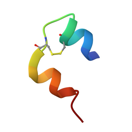
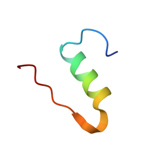
    We previously introduced the spitrobot, a protein crystal plunging system that enables reaction quenching via cryo-trapping with a time resolution in the millisecond range. Here we present the next generation, spitrobot-2, as an integrated benchtop device. User-friendliness has been improved by semi-automatic sample exchange. Moreover, a fully automated shutter shields the liquid nitrogen from the humidified environment, improving sample integrity. Most importantly, the cryo-trapping delay time has been reduced to 23 ms, making spitrobot-2 twice as fast as the previous generation. This further expands the number of target systems that can be addressed by cryo-trapping time-resolved crystallography. Using 12 crystal structures of three independent model systems, we demonstrate successful cryo-trapping via observation of conformational changes and ligand binding within 25 ms. These improvements increase the convenient access to cryo-trapping, time-resolved X-ray crystallography empowering the MX community with efficient tools to advance research in structural biology.


  • Organizational Affiliation
    • University Medical Center Hamburg-Eppendorf (UKE), Hamburg, Germany.

Macromolecule Content 

  • Total Structure Weight: 5.91 kDa 
  • Atom Count: 503 
  • Modeled Residue Count: 51 
  • Deposited Residue Count: 51 
  • Unique protein chains: 2

Macromolecules

Find similar proteins by:  Sequence   |   3D Structure  
Entity ID: 1
MoleculeChains  Sequence LengthOrganismDetailsImage
Insulin A chain21Homo sapiensMutation(s): 0 
Gene Names: INS
UniProt & NIH Common Fund Data Resources
Find proteins for P01308 (Homo sapiens)
Explore P01308 
Go to UniProtKB:  P01308
PHAROS:  P01308
GTEx:  ENSG00000254647 
Entity Groups
Sequence Clusters30% Identity50% Identity70% Identity90% Identity95% Identity100% Identity
UniProt GroupP01308
Sequence Annotations
Expand
Reference Sequence
Find similar proteins by:|  3D Structure
Entity ID: 2
MoleculeChains  Sequence LengthOrganismDetailsImage
Insulin B chain30Homo sapiensMutation(s): 0 
Gene Names: INS
UniProt & NIH Common Fund Data Resources
Find proteins for P01308 (Homo sapiens)
Explore P01308 
Go to UniProtKB:  P01308
PHAROS:  P01308
GTEx:  ENSG00000254647 
Entity Groups
Sequence Clusters30% Identity50% Identity70% Identity90% Identity95% Identity100% Identity
UniProt GroupP01308
Sequence Annotations
Expand
Reference Sequence

Small Molecules

Ligands 1 Unique
IDChains Name / Formula / InChI Key2D Diagram3D Interactions
SO4
(Subject of Investigation/LOI)

Query on SO4


Download:Ideal Coordinates CCD File
C [auth B]SULFATE ION
O4 S
QAOWNCQODCNURD-UHFFFAOYSA-L

Experimental Data & Validation

Experimental Data

  • Method: X-RAY DIFFRACTION
  • Resolution: 1.45 Å
  • R-Value Free:  0.196 (Depositor) 
  • R-Value Work:  0.170 (Depositor) 
  • R-Value Observed: 0.172 (Depositor) 
Space Group: I 21 3
Unit Cell:
Length ( Å )Angle ( ˚ )
a = 78.345α = 90
b = 78.345β = 90
c = 78.345γ = 90
Software Package:
Software NamePurpose
XDSdata reduction
autoPROCdata reduction
Aimlessdata scaling
autoPROCdata scaling
PHASERphasing
REFMACrefinement

Structure Validation

View Full Validation Report


Ligand Structure Quality Assessment 


Entry History 

& Funding Information

Deposition Data


Funding OrganizationLocationGrant Number
Max Planck SocietyGermany--
European Research Council (ERC)European Union--
German Research Foundation (DFG)Germany458246365
German Research Foundation (DFG)Germany451079909
German Federal Ministry for Education and ResearchGermany01KI2114

Revision History  (Full details and data files)

  • Version 1.0: 2025-12-03
    Type: Initial release