


Structural investigation of gain of function mutations in 1-phosphofructokinase (FruK) of E. coli
Dronsella, B., Zarzycki, J., Satanowski, A., Bar-Even, A., Erb, T.J., Lindner, S.N.To be published.
Experimental Data Snapshot
Entity ID: 1 | |||||
|---|---|---|---|---|---|
| Molecule | Chains | Sequence Length | Organism | Details | Image |
| 1-phosphofructokinase | 312 | Escherichia coli K-12 | Mutation(s): 1 Gene Names: fruK, fpk, b2168, JW2155 EC: 2.7.1.56 | 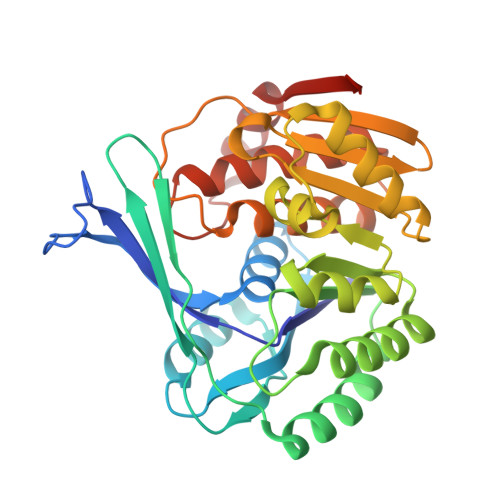 | |
UniProt | |||||
Entity Groups | |||||
| Sequence Clusters | 30% Identity50% Identity70% Identity90% Identity95% Identity100% Identity | ||||
| UniProt Group | P0AEW9 | ||||
Sequence AnnotationsExpand | |||||
Reference Sequence | |||||
| Ligands 6 Unique | |||||
|---|---|---|---|---|---|
| ID | Chains | Name / Formula / InChI Key | 2D Diagram | 3D Interactions | |
| ADP (Subject of Investigation/LOI) Download:Ideal Coordinates CCD File | E [auth A] | ADENOSINE-5'-DIPHOSPHATE C10 H15 N5 O10 P2 XTWYTFMLZFPYCI-KQYNXXCUSA-N |  | ||
| F1X (Subject of Investigation/LOI) Download:Ideal Coordinates CCD File | F [auth A] | 1-O-phosphono-beta-D-fructofuranose C6 H13 O9 P RHKKZBWRNHGJEZ-ARQDHWQXSA-N |  | ||
| GOL Download:Ideal Coordinates CCD File | G [auth A] | GLYCEROL C3 H8 O3 PEDCQBHIVMGVHV-UHFFFAOYSA-N |  | ||
| MG (Subject of Investigation/LOI) Download:Ideal Coordinates CCD File | B [auth A] | MAGNESIUM ION Mg JLVVSXFLKOJNIY-UHFFFAOYSA-N |  | ||
| NA Download:Ideal Coordinates CCD File | D [auth A] | SODIUM ION Na FKNQFGJONOIPTF-UHFFFAOYSA-N |  | ||
| LI Download:Ideal Coordinates CCD File | C [auth A] | LITHIUM ION Li HBBGRARXTFLTSG-UHFFFAOYSA-N |  | ||
| Length ( Å ) | Angle ( ˚ ) |
|---|---|
| a = 62.98 | α = 90 |
| b = 98.12 | β = 90 |
| c = 139.85 | γ = 90 |
| Software Name | Purpose |
|---|---|
| PHENIX | refinement |
| XSCALE | data scaling |
| XDS | data reduction |
| PHENIX | phasing |
| PDB_EXTRACT | data extraction |
| Funding Organization | Location | Grant Number |
|---|---|---|
| Max Planck Society | Germany | -- |
| German Federal Ministry for Education and Research | Germany | 033RC023G |


